ARG30267
Organelle Marker, Cytoplasm, Nucleus Antibody Duo (Histone, GAPDH)
内含物
| 货号 | 内含物名称 | 宿主克隆性 | 反应 | 应用 | 包装 |
|---|---|---|---|---|---|
| ARG65681 | anti-Histone H3 antibody | Mouse mAb | Hu, Ms, Rat | ICC/IF, IHC-P, IP, WB | 50 μg |
| ARG10112 | anti-GAPDH antibody [6C5] | Mouse mAb | AGMK, Bb, Cat, Chk, Dog, Fsh, Hm, Hu, Mk, Ms, Pig, Rb, Rat, Xenopus laevis, Zfsh | ELISA, ICC/IF, IHC-Fr, WB | 50 μg |
概述
| 产品描述 | GAPDH is often stably and constitutively expressed at high levels in most tissues and cells in cytoplasm. Therefore, GAPDH is a widely used loading control and cytoplasm marker for western blotting (35-40kDa) and Immunostaining study. Histone H3 (15-20kDa) is one of the component of nucleosomes in nucleus. So Histone H3 is one of the commonly used loading control and nucleus marker in western blotting and Immunostaining study. This Cytoplasm, Nucleus antibody Duos not only provide different MW targets for internal control selection, but also provide cytoplasm (GAPDH) and nucleus (Histone H3) markers to control the quality of cytoplasm and nucleus fractions after subcellular fractionation process and to identify the cytoplasm and nucleus position in Immunostaining study. |
|---|---|
| 靶点名称 | Organelle Marker, Cytoplasm, Nucleus |
| 別名 | Organelle Marker, Cytoplasm, Nucleus antibody; GAPDH antibody; Histone H3 antibody |
属性
| 存放说明 | For continuous use, store undiluted antibody at 2-8°C for up to a week. For long-term storage, aliquot and store at -20°C or below. Storage in frost free freezers is not recommended. Avoid repeated freeze/thaw cycles. Suggest spin the vial prior to opening. The antibody solution should be gently mixed before use. |
|---|---|
| 注意事项 | For laboratory research only, not for drug, diagnostic or other use. |
生物信息
| 全名 | Antibody Duo for Organelle Marker, Cytoplasm, Nucleus (Histone, GAPDH) |
|---|---|
| 产品亮点 | Related Product: anti-Histone H3 antibody; anti-GAPDH antibody; |
检测图片 (19) Click the Picture to Zoom In
-

ARG65681 anti-Histone H3 antibody ICC/IF image
Immunofluorescence: 100% Methanol fixed (RT, 10 min) HeLa cells stained with ARG65681 anti-Histone H3 antibody at 1:100 dilution. Left: primary antibody (green). Right: Merge (primary antibody and DAPI).
Secondary antibody: ARG55393 Goat anti-Mouse IgG (H+L) antibody (FITC)
-
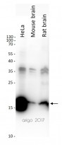
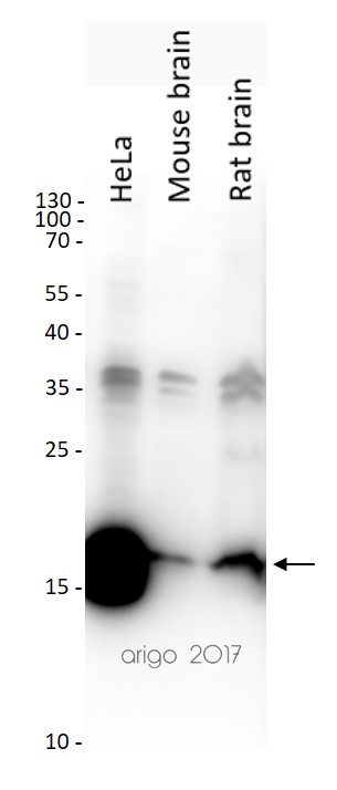
ARG65681 anti-Histone H3 antibody WB image
Western blot: 20 µg of HeLa, Mouse brain and Rat brain lysates stained with ARG65681 anti-Histone H3 antibody at 1:2000 dilution.
-
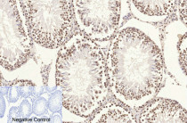
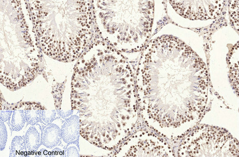
ARG65681 anti-Histone H3 antibody IHC-P image
Immunohistochemistry: Paraffin-embedded Rat testis tissue stained with ARG65681 anti-Histone H3 antibody at 1:200 dilution (4°C, overnight). Antigen Retrieval: Boil tissue section in Sodium citrate buffer (pH 6.0) for 20 min. Secondary antibody was diluted at 1:200 (RT, 30 min).
Negative control was used by secondary antibody only.
-

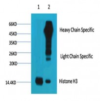
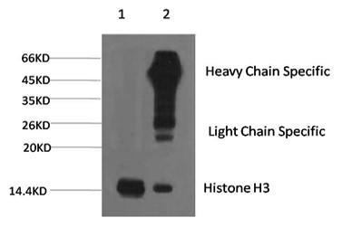
ARG65681 anti-Histone H3 antibody IP image
Immunoprecipitation: 1) HeLa cell lysate stained with ARG65681 anti-Histone H3 antibody and 2) IP product immunoprecipitated by ARG65681 anti-Histone H3 antibody at 1:200 dilution.
-
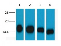
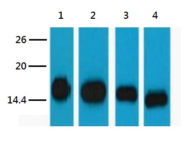
ARG65681 anti-Histone H3 antibody WB image
Western blot: 1) HeLa, 2) Raw, 3) Mouse brain tissue, and 4) Rat brain tissue lysates stained with ARG65681 anti-Histone H3 antibody at 1:5000 dilution.
-
ARG65681 anti-Histone H3 antibody IP image
Immunoprecipitation: 1) HeLa cell lysate stained with ARG65681 anti-Histone H3 antibody and 2) IP product immunoprecipitated by ARG65681 anti-Histone H3 antibody at 1:200 dilution.
-
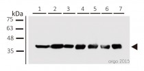
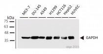
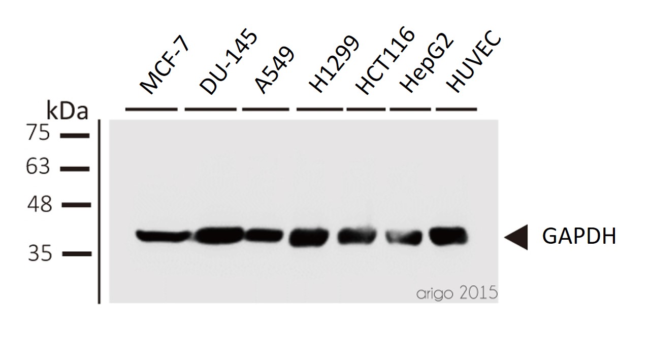
ARG10112 anti-GAPDH antibody [6C5] WB image
Western blot: 1) MCF-7 2) DU-145 3) A549 4) H1299 5) HCT116 6) HepG2 7) HUVEC stained with ARG10112 anti-GAPDH antibody [6C5] at 1:1000 dilution.
-
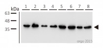
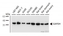
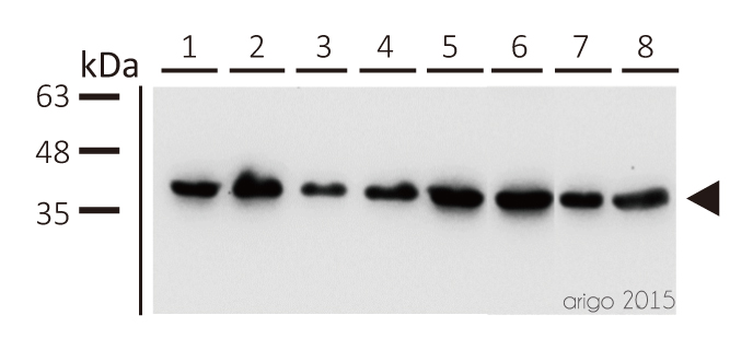
ARG10112 anti-GAPDH antibody [6C5] WB image
Western blot: 1) U87-MG 2) MCF-7 3) A549 4) DU145 5) SW480 6) rat brain 7) rat stomach 8) rat ovary stained with ARG10112 anti-GAPDH antibody [6C5] at 1:5000 dilution.
-

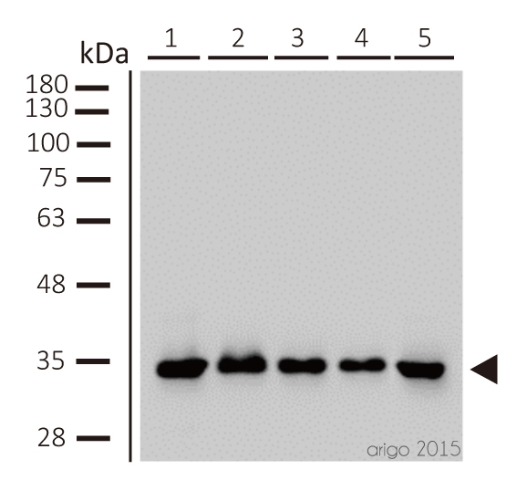
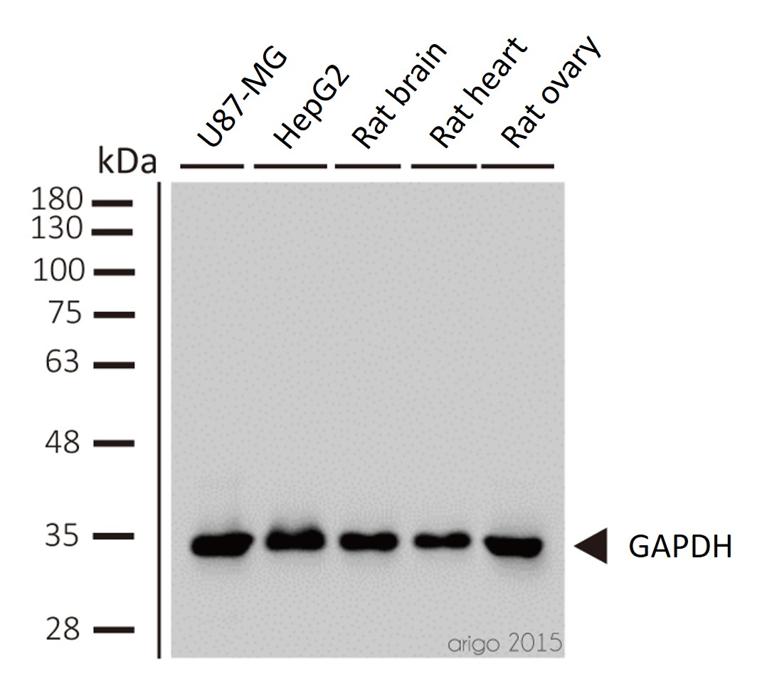
ARG10112 anti-GAPDH antibody [6C5] WB image
Western blot: 1) U87-MG 2) HepG2 3) rat brain 4) rat heart 5) rat ovary stained with ARG10112 anti-GAPDH antibody [6C5] at 1:2000 dilution.
-

ARG10112 anti-GAPDH antibody [6C5] WB image
Western blot: pAFs, AcoSV40, and AcoSI-1 stained with ARG10112 anti-GAPDH antibody [6C5] at 1:5000 dilution.
From Michele N Dill et al. PLoS One. (2023), doi: 10.3389/fcell.2022.899869, Fig. 2. C.
-

ARG10112 anti-GAPDH antibody [6C5] WB image
Western blot: Porcine kidney stained with ARG10112 anti-GAPDH antibody [6C5].
From Jianni Huang et al. Front Cell Dev Biol (2022), doi: 10.3389/fcell.2022.899869, Fig. 2. E.
-
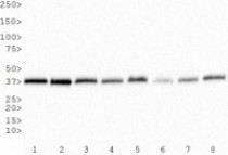
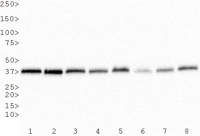
ARG10112 anti-GAPDH antibody [6C5] WB image
Western Blot: 1) HeLa, 2) NTERA-2, 3) A-431, 4) HepG2, 5) MCF-7, 6) NIH 3T3, 7) PC-12 and 8) COS-7 whole cell lysates stained with anti-GAPDH antibody [6C5] (ARG10112)
-

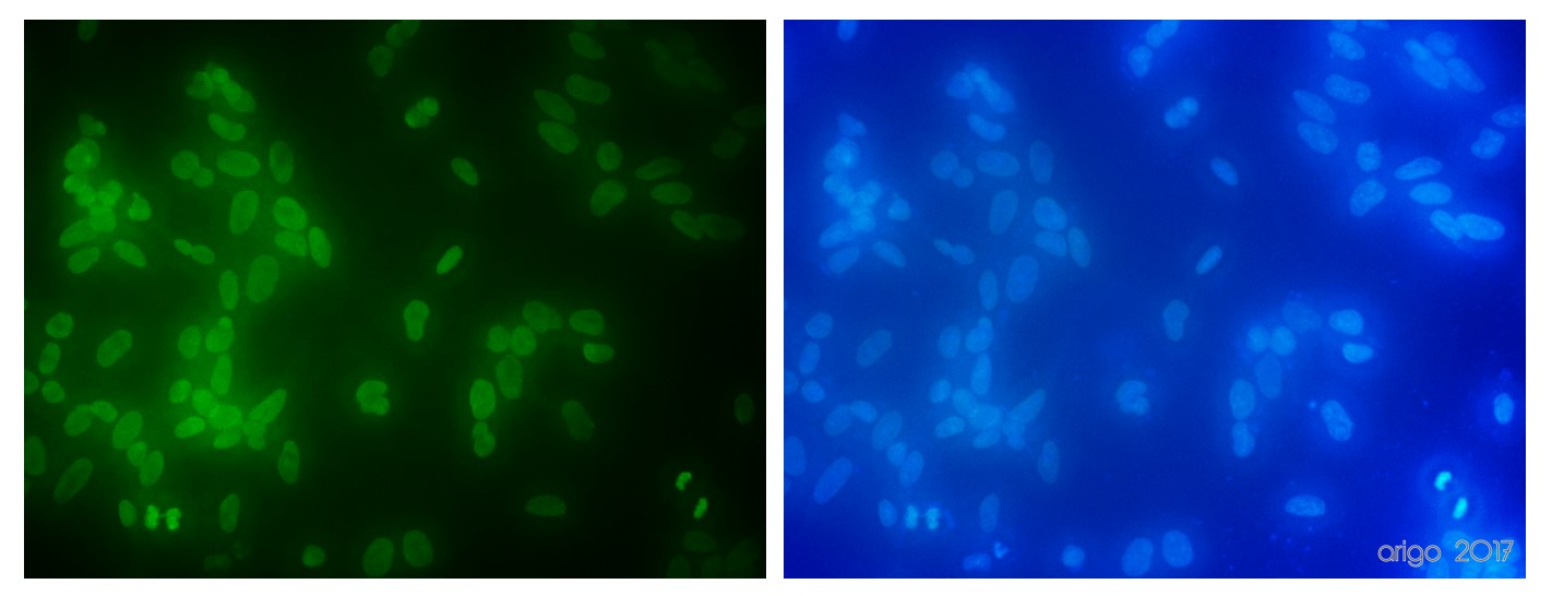
ARG10112 anti-GAPDH antibody [6C5] ICC/IF image
Immunofluorescence: 100% Methanol fixed (RT, 10 min) HeLa cells stained with ARG10112 anti-GAPDH antibody [6C5] (green) at 1:200 dilution.
Secondary antibody: ARG55393 Goat anti-Mouse IgG (H+L) antibody (FITC)
-
ARG10112 anti-GAPDH antibody [6C5] WB image
Western blot: Mouse samples stained with ARG10112 anti-GAPDH antibody [6C5] at 1:1000 dilution.
From Yun-Yun Li et al. Int J Biol Sci (2022), doi: 10.7150/ijbs.68224, Fig. 5. C.
-
ARG10112 anti-GAPDH antibody [6C5] WB image
Western blot: 1) MCF-7 2) DU-145 3) A549 4) H1299 5) HCT116 6) HepG2 7) HUVEC stained with ARG10112 anti-GAPDH antibody [6C5] at 1:1000 dilution.
-

ARG10112 anti-GAPDH antibody [6C5] WB image
Western blot: 1) U87-MG 2) HepG2 3) rat brain 4) rat heart 5) rat ovary stained with ARG10112 anti-GAPDH antibody [6C5] at 1:2000 dilution.
-

ARG10112 anti-GAPDH antibody [6C5] WB image
Western blot: HUVEC stained with ARG10112 anti-GAPDH antibody [6C5].
From Bingzheng Lu et al. Oxid Med Cell Longev (2020), doi: 10.1155/2020/2048210, Fig. 5. B.
-
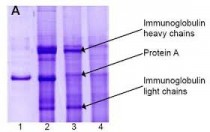
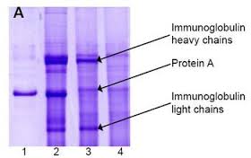
ARG10112 anti-GAPDH antibody [6C5] IP image
Immunoprecipitation and western blot: 1) GAPDH (1 μg). 2) GAPDH IP from rat heart tissue extract. 3) Only GAPDH preincubated with Protein A Sepharose. 4) Only Protein A Sepharose stained with ARG10112 GAPDH antibody [6C5].
Mixture of protein A-Sepharose with ARG10112 anti-GAPDH and tissue extract was incubated for 30 min at room temperature and precipitated by centrifugation. Pellet was washed with PBS, suspended in reducing electrophoresis sample buffer and heated for 5 minutes at 100ºC. After centrifugation supernatant was loaded on gel and proteins were separated by SDS electrophoresis. -
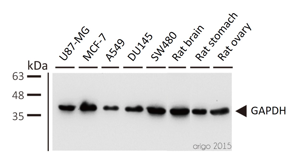
ARG10112 anti-GAPDH antibody [6C5] WB image
Western blot: 1) U87-MG 2) MCF-7 3) A549 4) DU145 5) SW480 6) rat brain 7) rat stomach 8) rat ovary stained with ARG10112 anti-GAPDH antibody [6C5] at 1:5000 dilution.


文献引用
Alleviation of liver fibrosis by inhibiting a non-canonical ATF4-regulated enhancer program in hepatic stellate cells
ARG10112: WB / Human


KIF9 Ameliorates Neuropathology and Cognitive Dysfunction by Promoting Macroautophagy in a Mouse Model of Alzheimer's Disease
ARG10112: WB / Human, Mouse
The effect of long-term administration of green tea catechins on aging-related cardiac diastolic dysfunction and decline of troponin I
ARG10112: WB / Mouse
PCSK6 exacerbates Alzheimer's disease pathogenesis by promoting MT5-MMP maturation
ARG10112: WB / Human, Mouse
Biochanin A inhibits cardiac hypertrophy and fibrosis in vivo and in vitro
ARG10112: WB / Mouse, Rat
Shionone relieves oxygen-glucose deprivation/reoxygenation induced SH-SY5Y cells injury by inhibiting the p38 MAPK/NF-κB pathway
ARG10112: WB / Human
7,8-Dihydroxyflavone ameliorates cognitive impairment induced by repeated neonatal sevoflurane exposures in mice through increasing tau O-GlcNAcylation
ARG10112: WB / Mouse
黄芪甲苷预处理对大鼠肠缺血再灌注所致肺损伤的影响及其机制
ARG10112: WB / Rat
PPARγ activation suppresses chondrocyte ferroptosis through mitophagy in osteoarthritis
ARG10112: WB / Rat
Discovery of the First Subnanomolar PPARα/δ Dual Agonist for the Treatment of Cholestatic Liver Diseases
ARG10112: WB / Mouse
Knockdown of Porf-2 restores visual function after optic nerve crush injury
ARG10112: WB / Mouse
Resveratrol inhibits Toxoplasma gondii-induced lung injury, inflammatory cascade and evidences of its mechanism of action
ARG10112: WB / Mouse
Uric acid mitigates cognitive deficits via transcription factor EB - mediated microglial autophagy in mice models of Alzheimer's disease
ARG10112: WB / Mouse
SCM-198 Prevents Endometriosis by Reversing Low Autophagy of Endometrial Stromal Cell via Balancing ERα and PR Signals
ARG10112: WB / Mouse
Pharmacological inhibition of SMYD2 protects against cisplatin-induced acute kidney injury in mice
ARG10112: WB / Mouse
Mesenchymal Stem Cells-Derived Exosomes Ameliorate Ischemia/Reperfusion Induced Acute Kidney Injury in a Porcine Model
ARG10112: WB / Pig
Lack of interferon regulatory factor 3 leads to anxiety/depression-like behaviors through disrupting the balance of neuronal excitation and inhibition in mice
ARG10112: WB / Mouse
Downregulation of lncRNA NEAT1 alleviates sepsis-induced acute kidney injury
ARG10112: WB / Human
Genetic labeling reveals cellular expression pattern of neuregulin 1 in mouse forebrain
ARG10112: WB / Human
Qi-Tai-Suan, an oleanolic acid derivative, ameliorates ischemic heart failure via suppression of cardiac apoptosis, inflammation and fibrosis
ARG10112: WB / Human
Gut microbiome dysbiosis contributes to abdominal aortic aneurysm by promoting neutrophil extracellular trap formation
ARG65681: IHC-P / Mouse
M351-0056 is a novel low MW compound modulating the actions of the immune-checkpoint protein VISTA.
ARG10112: WB / Human, Mouse
7,8-dihydroxyflavone ameliorates motor deficits via regulating autophagy in MPTP-induced mouse model of Parkinson's disease
ARG10112: WB / Mouse
Cardiac-Specific Gene TNNI3 as a Potential Oncogene for Kidney Cancer and Its Involvement in Wnt Signaling Pathway.
ARG10112: WB / Human
Mesenchymal Stem Cell-Derived Exosomes Ameliorate Alzheimer's Disease Pathology and Improve Cognitive Deficits.
ARG10112: WB / Human
Circ_0081572 inhibits the progression of periodontitis through regulating the miR-378h/RORA axis.
ARG10112: WB / Human
Evaluation of Class IIa Histone Deacetylases Expression and In Vivo Epigenetic Imaging in a Transgenic Mouse Model of Alzheimer's Disease
ARG10112: WB / Human
Structural Basis of VSIG3: The Ligand for VISTA.
ARG10112: WB / Human
The Phosphorylation Status of Drp1-Ser637 by PKA in Mitochondrial Fission Modulates Mitophagy via PINK1/Parkin to Exert Multipolar Spindles Assembly during Mitosis.
ARG10112: WB / Human
Curcumin attenuates renal ischemia reperfusion injury via JNK pathway with the involvement of p300/CBP-mediated histone acetylation
ARG10112: WB / Rat


