ARG66247
anti-Bax antibody [SQab1736]
anti-Bax antibody [SQab1736] for Flow cytometry,IHC-Formalin-fixed paraffin-embedded sections,Immunoprecipitation,Western blot and Human,Mouse,Rat
Cancer antibody; Cell Biology and Cellular Response antibody; Cell Death antibody; Metabolism antibody; Mitochondrial fission antibody; Apoptosis Marker antibody; Pro-apoptotic Bcl2 protein antibody
概述
| 产品描述 | Recombinant Rabbit Monoclonal antibody [SQab1736] recognizes Bax |
|---|---|
| 反应物种 | Hu, Ms, Rat |
| 应用 | FACS, IHC-P, IP, WB |
| 宿主 | Rabbit |
| 克隆 | Monoclonal |
| 克隆号 | SQab1736 |
| 同位型 | IgG |
| 靶点名称 | Bax |
| 抗原物种 | Human |
| 抗原 | Synthetic peptide around aa. 1-100 (C-terminus) of Human Bax. |
| 偶联标记 | Un-conjugated |
| 別名 | Bcl-2-like protein 4; Bcl2-L-4; BCL2L4; Apoptosis regulator BAX |
应用说明
| 应用建议 |
|
||||||||||
|---|---|---|---|---|---|---|---|---|---|---|---|
| 应用说明 | IHC-P: Antigen Retrieval: Boil tissue section in Tris/EDTA buffer (pH 9.0). * The dilutions indicate recommended starting dilutions and the optimal dilutions or concentrations should be determined by the scientist. |
属性
| 形式 | Liquid |
|---|---|
| 纯化 | Purification with Protein A. |
| 缓冲液 | PBS, 0.01% Sodium azide, 40% Glycerol and 0.05% BSA. |
| 抗菌剂 | 0.01% Sodium azide |
| 稳定剂 | 40% Glycerol and 0.05% BSA |
| 存放说明 | For continuous use, store undiluted antibody at 2-8°C for up to a week. For long-term storage, aliquot and store at -20°C. Storage in frost free freezers is not recommended. Avoid repeated freeze/thaw cycles. Suggest spin the vial prior to opening. The antibody solution should be gently mixed before use. |
| 注意事项 | For laboratory research only, not for drug, diagnostic or other use. |
生物信息
| 数据库连接 | |
|---|---|
| 基因名称 | BAX |
| 全名 | BCL2-associated X protein |
| 背景介绍 | Bax belongs to the BCL2 protein family. BCL2 family members form hetero- or homodimers and act as anti- or pro-apoptotic regulators that are involved in a wide variety of cellular activities. This protein forms a heterodimer with BCL2, and functions as an apoptotic activator. The association and the ratio of BAX to BCL2 also determines survival or death of a cell following an apoptotic stimulus. This protein is reported to interact with, and increase the opening of, the mitochondrial voltage-dependent anion channel (VDAC), which leads to the loss in membrane potential and the release of cytochrome c. The expression of this gene is regulated by the tumor suppressor P53 and has been shown to be involved in P53-mediated apoptosis. Multiple alternatively spliced transcript variants, which encode different isoforms, have been reported for this gene. [provided by RefSeq, Dec 2019] |
| 生物功能 | Bax plays a role in the mitochondrial apoptotic process. Under normal conditions, BAX is largely cytosolic via constant retrotranslocation from mitochondria to the cytosol mediated by BCL2L1/Bcl-xL, which avoids accumulation of toxic BAX levels at the mitochondrial outer membrane (MOM) (PubMed:21458670). Under stress conditions, undergoes a conformation change that causes translocation to the mitochondrion membrane, leading to the release of cytochrome c that then triggers apoptosis. Promotes activation of CASP3, and thereby apoptosis. [UniProt] |
| 产品亮点 | Related products: Bax antibodies; Bax Duos / Panels; Anti-Rabbit IgG secondary antibodies; Related news: Cancer Pathology Markers (SQ clones) |
| 研究领域 | Cancer antibody; Cell Biology and Cellular Response antibody; Cell Death antibody; Metabolism antibody; Mitochondrial fission antibody; Apoptosis Marker antibody; Pro-apoptotic Bcl2 protein antibody |
| 预测分子量 | 21 kDa |
检测图片 (6) Click the Picture to Zoom In
-
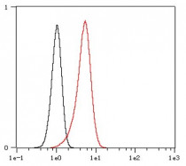
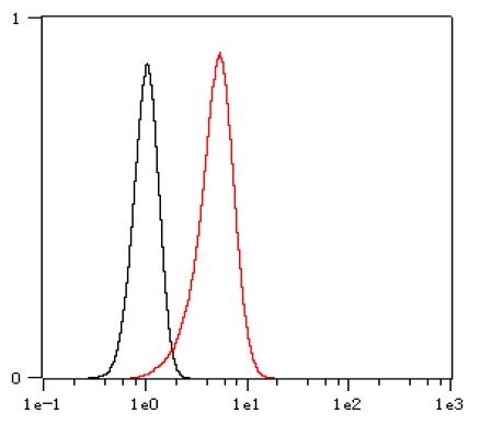
ARG66247 anti-Bax antibody [SQab1736] FACS image
Flow Cytometry: HeLa cells were fixed with 4% paraformaldehyde (10 min) and then permeabilized with 0.1% TritonX-100 for 15 min. The cells were then stained with ARG66247 anti-Bax antibody [SQab1736] (red) at 1:400 dilution in 1x PBS/1% BSA for 30 min at room temperature, followed by Alexa Fluor® 488 labelled secondary antibody. Unlabelled sample (black) was used as a control.
-
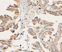
ARG66247 anti-Bax antibody [SQab1736] IHC-P image
Immunohistochemistry: Formalin-fixed and paraffin-embedded Ovarian cancer tissue stained with ARG66247 anti-Bax antibody [SQab1736] at 1:1600. Antigen Retrieval: Boil tissue section in Tris/EDTA buffer (pH 9.0).
-
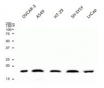
ARG66247 anti-Bax antibody [SQab1736] WB image
Western blot: 10 µg of OVCAR-3, A549, HT-29, SH-SY5Y and LnCap cell lysates stained with ARG66247 anti-Bax antibody [SQab1736] at 1:1000 dilution.
-
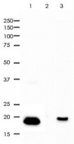
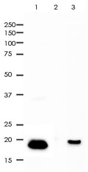
ARG66247 anti-Bax antibody [SQab1736] IP image
Immunoprecipitation: BAX was immunioprecipitated from 0.5 mg HT-29 whole cell lysate with ARG66247 anti-Bax antibody [SQab1736] at 1:25 dilution. Western blot was performed from the immunoprecipitate using ARG66247 anti-Bax antibody [SQab1736] at 1:2000 dilution.
1. IP by using ARG66247 in HT-29 whole cell lysate
2. PBS instead of ARG66247 in HT-29 whole cell lysate
3. 10 µg of HT-29 whole cell lysate (input) -

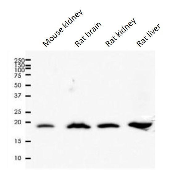
ARG66247 anti-Bax antibody [SQab1736] WB image
Western blot: 10 µg of Mouse kidney, Rat brain, Rat kidney and Rat liver lysates stained with ARG66247 anti-Bax antibody [SQab1736] at 1:1000 dilution.
-

ARG66247 anti-Bax antibody [SQab1736] WB image
Western blot: Gastric cancer cells stained with ARG66247 anti-Bax antibody [SQab1736].
From Limin Zhang et al. Heliyon (2024), doi: 10.1016/j.heliyon.2024.e30803, Fig. 4. C.


文献引用
Suppression of gastric cancer cell proliferation by miR-494-3p inhibitor-loaded engineered exosomes
WB / Human


Resveratrol improves ovarian state by inhibiting apoptosis of granulosa cells
WB / Human
Apoptosis inhibition is involved in improvement of sevoflurane-induced cognitive impairment following normobaric hyperoxia preconditioning in aged rats.
WB / Rat
Circ_0081572 inhibits the progression of periodontitis through regulating the miR-378h/RORA axis.
WB / Human
miR-371b-5p promotes cell proliferation, migration and invasion in non-small cell lung cancer via SCAI
WB / Human


