ARG42699
anti-DHX15 / Prp43 antibody
anti-DHX15 / Prp43 antibody for Flow cytometry,ICC/IF,IHC-Formalin-fixed paraffin-embedded sections,Western blot and Human,Mouse,Rat
概述
| 产品描述 | Rabbit Polyclonal antibody recognizes DHX15 / Prp43 |
|---|---|
| 反应物种 | Hu, Ms, Rat |
| 应用 | FACS, ICC/IF, IHC-P, WB |
| 宿主 | Rabbit |
| 克隆 | Polyclonal |
| 同位型 | IgG |
| 靶点名称 | DHX15 / Prp43 |
| 抗原物种 | Human |
| 抗原 | Synthetic peptide corresponding to a sequence of Human DHX15 / Prp43. (DIKPEWLVKIAPQYYDMSNFPQCEAKRQLDRIIAKLQSKEYSQY) |
| 偶联标记 | Un-conjugated |
| 別名 | Pre-mRNA-splicing factor ATP-dependent RNA helicase DHX15; DDX15; PrPp43p; ATP-dependent RNA helicase #46; PRP43; EC 3.6.4.13; DEAH box protein 15; HRH2; DBP1; PRPF43 |
应用说明
| 应用建议 |
|
||||||||||
|---|---|---|---|---|---|---|---|---|---|---|---|
| 应用说明 | * The dilutions indicate recommended starting dilutions and the optimal dilutions or concentrations should be determined by the scientist. | ||||||||||
| 实际分子量 | ~ 91 kDa |
属性
| 形式 | Liquid |
|---|---|
| 纯化 | Affinity purification with immunogen. |
| 缓冲液 | 0.2% Na2HPO4, 0.9% NaCl, 0.05% Sodium azide and 4% Trehalose. |
| 抗菌剂 | 0.05% Sodium azide |
| 稳定剂 | 4% Trehalose |
| 浓度 | 0.5 mg/ml |
| 存放说明 | For continuous use, store undiluted antibody at 2-8°C for up to a week. For long-term storage, aliquot and store at -20°C or below. Storage in frost free freezers is not recommended. Avoid repeated freeze/thaw cycles. Suggest spin the vial prior to opening. The antibody solution should be gently mixed before use. |
| 注意事项 | For laboratory research only, not for drug, diagnostic or other use. |
生物信息
| 数据库连接 |
Swiss-port # O35286 Mouse Pre-mRNA-splicing factor ATP-dependent RNA helicase DHX15 Swiss-port # O43143 Human Pre-mRNA-splicing factor ATP-dependent RNA helicase DHX15 |
|---|---|
| 基因名称 | DHX15 |
| 全名 | DEAH (Asp-Glu-Ala-His) box helicase 15 |
| 背景介绍 | The protein encoded by this gene is a putative ATP-dependent RNA helicase implicated in pre-mRNA splicing. [provided by RefSeq, Jul 2008] |
| 生物功能 | Pre-mRNA processing factor involved in disassembly of spliceosomes after the release of mature mRNA. In cooperation with TFIP11 seem to be involved in the transition of the U2, U5 and U6 snRNP-containing IL complex to the snRNP-free IS complex leading to efficient debranching and turnover of excised introns. [UniProt] |
| 细胞定位 | Nucleus. Nucleus, nucleolus. [UniProt] |
| 预测分子量 | 91 kDa |
检测图片 (11) Click the Picture to Zoom In
-
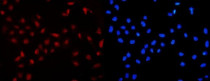
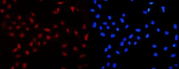
ARG42699 anti-DHX15 / Prp43 antibody ICC/IF image
Immunofluorescence: U2OS cells were blocked with 10% goat serum and then stained with ARG42699 anti-DHX15 / Prp43 antibody (red) at 2 µg/ml dilution, overnight at 4°C. DAPI (blue) for nuclear staining.
-
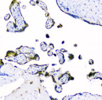
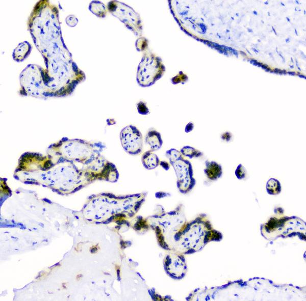
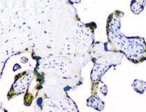
ARG42699 anti-DHX15 / Prp43 antibody IHC-P image
Immunohistochemistry: Paraffin-embedded Human placenta tissue. Antigen Retrieval: Heat mediation was performed in EDTA buffer (pH 8.0). The tissue section was blocked with 10% goat serum. The tissue section was then stained with ARG42699 anti-DHX15 / Prp43 antibody at 1 µg/ml dilution, overnight at 4°C.
-
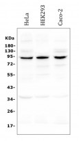
ARG42699 anti-DHX15 / Prp43 antibody WB image
Western blot: 50 µg of samples under reducing condition. HeLa, HEK293 and Caco-2 whole cell lysates stained with ARG42699 anti-DHX15 / Prp43 antibody at 0.5 µg/ml dilution, overnight at 4°C.
-
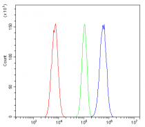
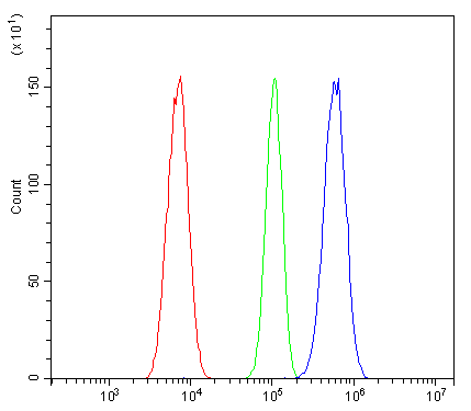
ARG42699 anti-DHX15 / Prp43 antibody FACS image
Flow Cytometry: A431 cells were blocked with 10% normal goat serum and then stained with ARG42699 anti-DHX15 / Prp43 antibody (blue) at 1 µg/10^6 cells for 30 min at 20°C, followed by incubation with DyLight®488 labelled secondary antibody. Isotype control antibody (green) was rabbit IgG (1 µg/10^6 cells) used under the same conditions. Unlabelled sample (red) was also used as a control.
-
ARG42699 anti-DHX15 / Prp43 antibody IHC-P image
Immunohistochemistry: Paraffin-embedded Human placenta tissue. Antigen Retrieval: Heat mediation was performed in EDTA buffer (pH 8.0). The tissue section was blocked with 10% goat serum. The tissue section was then stained with ARG42699 anti-DHX15 / Prp43 antibody at 1 µg/ml dilution, overnight at 4°C.
-
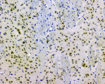
ARG42699 anti-DHX15 / Prp43 antibody IHC-P image
Immunohistochemistry: Paraffin-embedded Rat brain tissue. Antigen Retrieval: Heat mediation was performed in EDTA buffer (pH 8.0). The tissue section was blocked with 10% goat serum. The tissue section was then stained with ARG42699 anti-DHX15 / Prp43 antibody at 1 µg/ml dilution, overnight at 4°C.
-
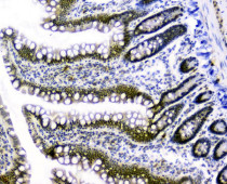
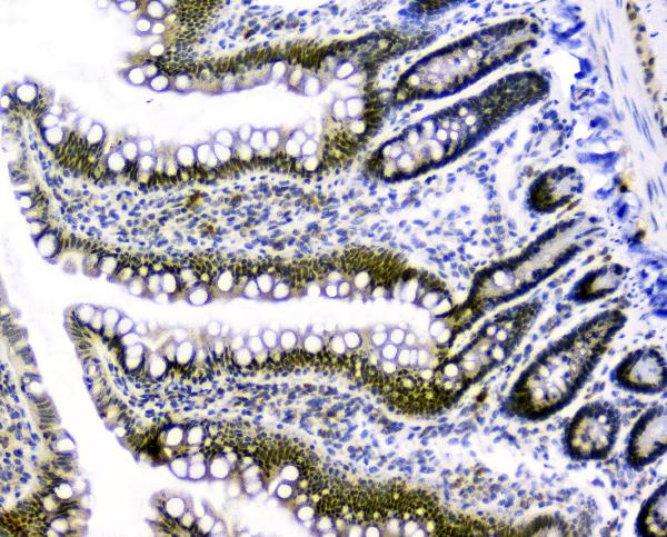
ARG42699 anti-DHX15 / Prp43 antibody IHC-P image
Immunohistochemistry: Paraffin-embedded Rat small intestine tissue. Antigen Retrieval: Heat mediation was performed in EDTA buffer (pH 8.0). The tissue section was blocked with 10% goat serum. The tissue section was then stained with ARG42699 anti-DHX15 / Prp43 antibody at 1 µg/ml dilution, overnight at 4°C.
-
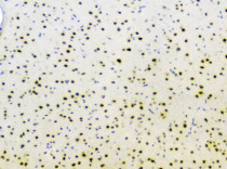
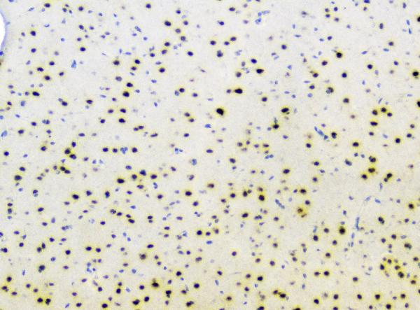
ARG42699 anti-DHX15 / Prp43 antibody IHC-P image
Immunohistochemistry: Paraffin-embedded Mouse brain tissue. Antigen Retrieval: Heat mediation was performed in EDTA buffer (pH 8.0). The tissue section was blocked with 10% goat serum. The tissue section was then stained with ARG42699 anti-DHX15 / Prp43 antibody at 1 µg/ml dilution, overnight at 4°C.
-
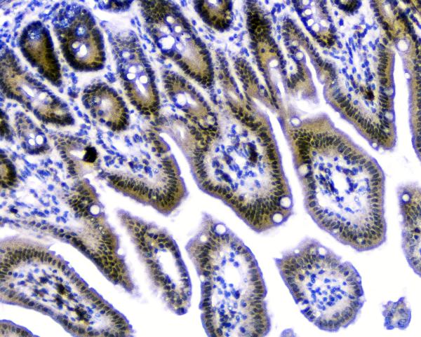
ARG42699 anti-DHX15 / Prp43 antibody IHC-P image
Immunohistochemistry: Paraffin-embedded Mouse small intestine tissue. Antigen Retrieval: Heat mediation was performed in EDTA buffer (pH 8.0). The tissue section was blocked with 10% goat serum. The tissue section was then stained with ARG42699 anti-DHX15 / Prp43 antibody at 1 µg/ml dilution, overnight at 4°C.
-
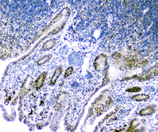
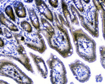
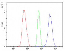
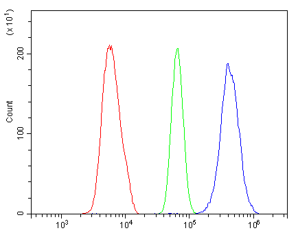
ARG42699 anti-DHX15 / Prp43 antibody FACS image
Flow Cytometry: ANA-1 cells were blocked with 10% normal goat serum and then stained with ARG42699 anti-DHX15 / Prp43 antibody (blue) at 1 µg/10^6 cells for 30 min at 20°C, followed by incubation with DyLight®488 labelled secondary antibody. Isotype control antibody (green) was rabbit IgG (1 µg/10^6 cells) used under the same conditions. Unlabelled sample (red) was also used as a control.