ARG55632
anti-HLA DR antibody [L243] (PE)
anti-HLA DR antibody [L243] (PE) for Flow cytometry and Human,Dog,Primates
Immune System antibody
概述
| 产品描述 | PE-conjugated Mouse Monoclonal antibody [L243] recognizes HLA DR |
|---|---|
| 反应物种 | Hu, Dog, NHuPrm |
| 应用 | FACS |
| 特异性 | This antibody recognizes specifically HLA-DR molecules, both peptide-loaded and empty. |
| 宿主 | Mouse |
| 克隆 | Monoclonal |
| 克隆号 | L243 |
| 同位型 | IgG2a |
| 靶点名称 | HLA DR |
| 抗原物种 | Human |
| 抗原 | Human B lymphocytes |
| 偶联标记 | PE |
| 別名 | MLRW; HLA class II histocompatibility antigen, DR alpha chain; HLA-DRA1; MHC class II antigen DRA |
应用说明
| 应用建议 |
|
||||
|---|---|---|---|---|---|
| 应用说明 | * The dilutions indicate recommended starting dilutions and the optimal dilutions or concentrations should be determined by the scientist. |
属性
| 形式 | Liquid |
|---|---|
| 缓冲液 | PBS and 15 mM Sodium azide |
| 抗菌剂 | 15 mM Sodium azide |
| 存放说明 | Aliquot and store in the dark at 2-8°C. Keep protected from prolonged exposure to light. Avoid repeated freeze/thaw cycles. Suggest spin the vial prior to opening. The antibody solution should be gently mixed before use. |
| 注意事项 | For laboratory research only, not for drug, diagnostic or other use. |
生物信息
| 数据库连接 |
Swiss-port # P01903 Human HLA class II histocompatibility antigen, DR alpha chain |
|---|---|
| 基因名称 | HLA-DRA |
| 全名 | major histocompatibility complex, class II, DR alpha |
| 背景介绍 | HLA-DRA is one of the HLA class II alpha chain paralogues. This class II molecule is a heterodimer consisting of an alpha and a beta chain, both anchored in the membrane. It plays a central role in the immune system by presenting peptides derived from extracellular proteins. Class II molecules are expressed in antigen presenting cells (APC: B lymphocytes, dendritic cells, macrophages). The alpha chain is approximately 33-35 kDa and its gene contains 5 exons. Exon 1 encodes the leader peptide, exons 2 and 3 encode the two extracellular domains, and exon 4 encodes the transmembrane domain and the cytoplasmic tail. DRA does not have polymorphisms in the peptide binding part and acts as the sole alpha chain for DRB1, DRB3, DRB4 and DRB5. [provided by RefSeq, Jul 2008] |
| 生物功能 | Binds peptides derived from antigens that access the endocytic route of antigen presenting cells (APC) and presents them on the cell surface for recognition by the CD4 T-cells. The peptide binding cleft accommodates peptides of 10-30 residues. The peptides presented by MHC class II molecules are generated mostly by degradation of proteins that access the endocytic route, where they are processed by lysosomal proteases and other hydrolases. Exogenous antigens that have been endocytosed by the APC are thus readily available for presentation via MHC II molecules, and for this reason this antigen presentation pathway is usually referred to as exogenous. As membrane proteins on their way to degradation in lysosomes as part of their normal turn-over are also contained in the endosomal/lysosomal compartments, exogenous antigens must compete with those derived from endogenous components. Autophagy is also a source of endogenous peptides, autophagosomes constitutively fuse with MHC class II loading compartments. In addition to APCs, other cells of the gastrointestinal tract, such as epithelial cells, express MHC class II molecules and CD74 and act as APCs, which is an unusual trait of the GI tract. To produce a MHC class II molecule that presents an antigen, three MHC class II molecules (heterodimers of an alpha and a beta chain) associate with a CD74 trimer in the ER to form a heterononamer. Soon after the entry of this complex into the endosomal/lysosomal system where antigen processing occurs, CD74 undergoes a sequential degradation by various proteases, including CTSS and CTSL, leaving a small fragment termed CLIP (class-II-associated invariant chain peptide). The removal of CLIP is facilitated by HLA-DM via direct binding to the alpha-beta-CLIP complex so that CLIP is released. HLA-DM stabilizes MHC class II molecules until primary high affinity antigenic peptides are bound. The MHC II molecule bound to a peptide is then transported to the cell membrane surface. In B-cells, the interaction between HLA-DM and MHC class II molecules is regulated by HLA-DO. Primary dendritic cells (DCs) also to express HLA-DO. Lysosomal microenvironment has been implicated in the regulation of antigen loading into MHC II molecules, increased acidification produces increased proteolysis and efficient peptide loading. [UniProt] |
| 研究领域 | Immune System antibody |
| 预测分子量 | 29 kDa |
| 翻译后修饰 | Ubiquitinated by MARCH1 or MARCH8 at Lys-244 leading to down-regulation of MHC class II. When associated with ubiquitination of the beta subunit of HLA-DR: HLA-DRB4 'Lys-254', the down-regulation of MHC class II may be highly effective. |
检测图片 (3) Click the Picture to Zoom In
-
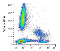
ARG55632 anti-HLA DR antibody [L243] (PE) FACS image
Flow Cytometry: Human peripheral whole blood stained with ARG55632 anti-HLA DR antibody [L243] (PE) (10 µl reagent / 100 µl of peripheral whole blood).
-
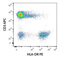
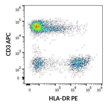
ARG55632 anti-HLA DR antibody [L243] (PE) FACS image
Flow Cytometry: Human lymphocytes stained with ARG54302 anti-CD3 antibody [UCHT1] (APC) (10 µl reagent / 100 µl of peripheral whole blood) and ARG55632 anti-HLA DR antibody [L243] (PE) (10 µl reagent / 100 µl of peripheral whole blood).
-
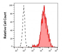
ARG55632 anti-HLA DR antibody [L243] (PE) FACS image
Flow Cytometry: Separation of human CD3 negative HLA-DR positive lymphocytes (red-filled) from neutrophil granulocytes (black-dashed). Human peripheral whole blood stained with ARG55632 anti-HLA DR antibody [L243] (PE) (10 µl reagent / 100 µl of peripheral whole blood).