ARG56033
anti-HLA DR antibody [LN3]
anti-HLA DR antibody [LN3] for Flow cytometry,ICC/IF,IHC-Formalin-fixed paraffin-embedded sections,Western blot and Human
概述
| 产品描述 | Mouse Monoclonal antibody [LN3] recognizes HLA DR |
|---|---|
| 反应物种 | Hu |
| 不反应物种 | Ms |
| 应用 | FACS, ICC/IF, IHC-P, WB |
| 宿主 | Mouse |
| 克隆 | Monoclonal |
| 克隆号 | LN3 |
| 同位型 | IgG2b, kappa |
| 靶点名称 | HLA DR |
| 抗原物种 | Human |
| 抗原 | Activated Human peripheral blood mononuclear cells. |
| 偶联标记 | Un-conjugated |
| 別名 | HLA-DRB; HLA class II histocompatibility antigen, DRB1-3 chain; SS1; MHC class II antigen DRB1*3; HLA-DR1B; DRw10; Clone P2-beta-3; DRB1 |
应用说明
| 应用建议 |
|
||||||||||
|---|---|---|---|---|---|---|---|---|---|---|---|
| 应用说明 | IHC-P: Antigen Retrieval: Boil tissue section in 10 mM Citrate buffer (pH 6.0) for 10-20 min, followed by cooling at RT for 20 min. * The dilutions indicate recommended starting dilutions and the optimal dilutions or concentrations should be determined by the scientist. |
属性
| 形式 | Liquid |
|---|---|
| 纯化 | Purification with Protein G. |
| 缓冲液 | PBS (pH 7.4), 0.05% Sodium azide and 0.1 mg/ml BSA |
| 抗菌剂 | 0.05% Sodium azide |
| 稳定剂 | 0.1 mg/ml BSA |
| 浓度 | 0.2 mg/ml |
| 存放说明 | For continuous use, store undiluted antibody at 2-8°C for up to a week. For long-term storage, aliquot and store at -20°C or below. Storage in frost free freezers is not recommended. Avoid repeated freeze/thaw cycles. Suggest spin the vial prior to opening. The antibody solution should be gently mixed before use. |
| 注意事项 | For laboratory research only, not for drug, diagnostic or other use. |
生物信息
| 数据库连接 |
Swiss-port # P01912 Human HLA class II histocompatibility antigen, DRB1-3 chain |
|---|---|
| 基因名称 | HLA-DRB1 |
| 全名 | major histocompatibility complex, class II, DR beta 1 |
| 背景介绍 | HLA-DRB1 belongs to the HLA class II beta chain paralogs. The class II molecule is a heterodimer consisting of an alpha (DRA) and a beta chain (DRB), both anchored in the membrane. It plays a central role in the immune system by presenting peptides derived from extracellular proteins. Class II molecules are expressed in antigen presenting cells (APC: B lymphocytes, dendritic cells, macrophages). The beta chain is approximately 26-28 kDa. It is encoded by 6 exons. Exon one encodes the leader peptide; exons 2 and 3 encode the two extracellular domains; exon 4 encodes the transmembrane domain; and exon 5 encodes the cytoplasmic tail. Within the DR molecule the beta chain contains all the polymorphisms specifying the peptide binding specificities. Hundreds of DRB1 alleles have been described and typing for these polymorphisms is routinely done for bone marrow and kidney transplantation. DRB1 is expressed at a level five times higher than its paralogs DRB3, DRB4 and DRB5. DRB1 is present in all individuals. Allelic variants of DRB1 are linked with either none or one of the genes DRB3, DRB4 and DRB5. There are 4 related pseudogenes: DRB2, DRB6, DRB7, DRB8 and DRB9. [provided by RefSeq, Jul 2008] |
| 生物功能 | Binds peptides derived from antigens that access the endocytic route of antigen presenting cells (APC) and presents them on the cell surface for recognition by the CD4 T-cells. The peptide binding cleft accommodates peptides of 10-30 residues. The peptides presented by MHC class II molecules are generated mostly by degradation of proteins that access the endocytic route; where they are processed by lysosomal proteases and other hydrolases. Exogenous antigens that have been endocytosed by the APC are thus readily available for presentation via MHC II molecules; and for this reason this antigen presentation pathway is usually referred to as exogenous. As membrane proteins on their way to degradation in lysosomes as part of their normal turn-over are also contained in the endosomal/lysosomal compartments; exogenous antigens must compete with those derived from endogenous components. Autophagy is also a source of endogenous peptides; autophagosomes constitutively fuse with MHC class II loading compartments. In addition to APCs; other cells of the gastrointestinal tract; such as epithelial cells; express MHC class II molecules and CD74 and act as APCs; which is an unusual trait of the GI tract. To produce a MHC class II molecule that presents an antigen; three MHC class II molecules (heterodimers of an alpha and a beta chain) associate with a CD74 trimer in the ER to form a heterononamer. Soon after the entry of this complex into the endosomal/lysosomal system where antigen processing occurs; CD74 undergoes a sequential degradation by various proteases; including CTSS and CTSL; leaving a small fragment termed CLIP (class-II-associated invariant chain peptide). The removal of CLIP is facilitated by HLA-DM via direct binding to the alpha-beta-CLIP complex so that CLIP is released. HLA-DM stabilizes MHC class II molecules until primary high affinity antigenic peptides are bound. The MHC II molecule bound to a peptide is then transported to the cell membrane surface. In B-cells; the interaction between HLA-DM and MHC class II molecules is regulated by HLA-DO. Primary dendritic cells (DCs) also to express HLA-DO. Lysosomal microenvironment has been implicated in the regulation of antigen loading into MHC II molecules; increased acidification produces increased proteolysis and efficient peptide loading. [UniProt] |
| 预测分子量 | 30 kDa |
| 翻译后修饰 | Ubiquitinated by MARCH1 and MARCH8 at Lys-254 leading to sorting into the endosome system and down-regulation of MHC class II. |
检测图片 (3) Click the Picture to Zoom In
-
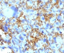
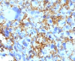
ARG56033 anti-HLA DR antibody [LN3] IHC-P image
Immunohistochemistry: Human histiocytoma stained with ARG56033 anti-HLA DR antibody [LN3].
-

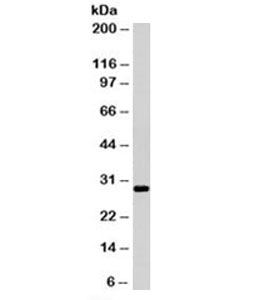
ARG56033 anti-HLA DR antibody [LN3] WB image
Western blot: Ramos cell lysate stained with ARG56033 anti-HLA DR antibody [LN3]. Expected molecular weight ~30kDa.
-

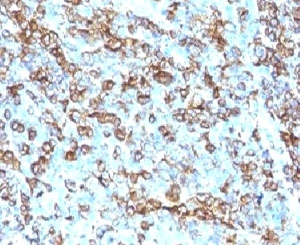
ARG56033 anti-HLA DR antibody [LN3] IHC-P image
Immunohistochemistry: Human tonsil stained with ARG56033 anti-HLA DR antibody [LN3].