ARG54707
anti-LC3B antibody
anti-LC3B antibody for ICC/IF,Immunohistochemistry,Western blot and Human,Mouse
Autophagy Study antibody

概述
| 产品描述 | Rabbit Polyclonal antibody recognizes LC3B |
|---|---|
| 反应物种 | Hu, Ms |
| 预测物种 | Bov |
| 应用 | ICC/IF, IHC, WB |
| 宿主 | Rabbit |
| 克隆 | Polyclonal |
| 同位型 | IgG |
| 靶点名称 | LC3B |
| 抗原物种 | Human |
| 抗原 | KLH-conjugated synthetic peptide around aa. 1-30 (N-terminus) of Human LC3 protein (NP_073729.1). |
| 偶联标记 | Un-conjugated |
| 別名 | Microtubule-associated proteins 1A/1B light chain 3B; MAP1A/MAP1B light chain 3 B; MAP1A/1BLC3; MAP1 light chain 3-like protein 2; Autophagy-related protein LC3 B; MAP1A/MAP1B LC3 B; LC3B; MAP1LC3B-a; ATG8F; Microtubule-associated protein 1 light chain 3 beta; Autophagy-related ubiquitin-like modifier LC3 B |
应用说明
| 应用建议 |
|
||||||||
|---|---|---|---|---|---|---|---|---|---|
| 应用说明 | * The dilutions indicate recommended starting dilutions and the optimal dilutions or concentrations should be determined by the scientist. |
属性
| 纯化 | This antibody is prepared by Saturated Ammonium Sulfate (SAS) precipitation followed by dialysis against PBS. |
|---|---|
| 缓冲液 | PBS and 0.09% (W/V) Sodium azide |
| 抗菌剂 | 0.09% (W/V) Sodium azide |
| 存放说明 | For continuous use, store undiluted antibody at 2-8°C for up to a week. For long-term storage, aliquot and store at -20°C or below. Storage in frost free freezers is not recommended. Avoid repeated freeze/thaw cycles. Suggest spin the vial prior to opening. The antibody solution should be gently mixed before use. |
| 注意事项 | For laboratory research only, not for drug, diagnostic or other use. |
生物信息
| 数据库连接 |
Swiss-port # Q9CQV6 Mouse Microtubule-associated proteins 1A/1B light chain 3B Swiss-port # Q9GZQ8 Human Microtubule-associated proteins 1A/1B light chain 3B |
|---|---|
| 基因名称 | MAP1LC3B |
| 全名 | microtubule-associated protein 1 light chain 3 beta |
| 背景介绍 | The product of this gene is a subunit of neuronal microtubule-associated MAP1A and MAP1B proteins, which are involved in microtubule assembly and important for neurogenesis. Studies on the rat homolog implicate a role for this gene in autophagy, a process that involves the bulk degradation of cytoplasmic component. [provided by RefSeq, Jul 2008] |
| 生物功能 | Ubiquitin-like modifier involved in formation of autophagosomal vacuoles (autophagosomes). Plays a role in mitophagy which contributes to regulate mitochondrial quantity and quality by eliminating the mitochondria to a basal level to fulfill cellular energy requirements and preventing excess ROS production. Whereas LC3s are involved in elongation of the phagophore membrane, the GABARAP/GATE-16 subfamily is essential for a later stage in autophagosome maturation. Promotes primary ciliogenesis by removing OFD1 from centriolar satellites via the autophagic pathway. [From Uniprot] |
| 细胞定位 | Cytoplasm, cytoskeleton. Endomembrane system; Lipid-anchor. Cytoplasmic vesicle, autophagosome membrane; Lipid-anchor. Note=LC3-II binds to the autophagic membranes Localizes also to discrete punctae along the ciliary axoneme (By similarity). |
| 产品亮点 | Related products: LC3B antibodies; LC3B Duos / Panels; Anti-Rabbit IgG secondary antibodies; Related news: Keap1-Nrf2-ARE antibody panel is launched |
| 研究领域 | Autophagy Study antibody |
| 翻译后修饰 | The precursor molecule is cleaved by ATG4B to form the cytosolic form, LC3-I. This is activated by APG7L/ATG7, transferred to ATG3 and conjugated to phospholipid to form the membrane-bound form, LC3-II (PubMed:15187094). The Legionella effector RavZ is a deconjugating enzyme that produces an ATG8 product that would be resistant to reconjugation by the host machinery due to the cleavage of the reactive C-terminal glycine. Phosphorylation at Thr-12 by PKA inhibits conjugation to phosphatidylethanolamine (PE) (By similarity). Interaction with MAPK15 reduces the inhibitory phosphorylation and increases autophagy activity. |
检测图片 (9) Click the Picture to Zoom In
-

ARG54707 anti-LC3B antibody WB image
Western blot: untreated or treated HeLa cell lysate stained with ARG54707 anti-LC3B antibody. Both non-lipidated (arrow, I) and lipidated LC3 (APG8b) (arrow, II) were detected. But pro-LC3 (APG8b) and non-lipidated LC3 (APG8b) were detected in soluble fraction (S).
-

ARG54707 anti-LC3B antibody WB image
Western blot: lysates from HepG2, mouse NIH/3T3 cell line, untreated or treated with 50uM chloroquine, stained with ARG54707 anti-LC3B antibody (upper) or Beta-actin (lower).
-

ARG54707 anti-LC3B antibody WB image
Western blot: HepG2 cell lysates untreated or treated with chloroquine stained with ARG54707 anti-LC3B antibody (upper) or Beta-actin (lower).
-

ARG54707 anti-LC3B antibody WB image
Western blot: NIH/3T3 cell lysates untreated or treated with chloroquine, stained with ARG54707 anti-LC3B antibody (upper) or Beta-actin (lower).
-

ARG54707 anti-LC3B antibody WB image
Western blot: rat brain lysate stained with ARG54707 anti-LC3B antibody. Both non-lipidated (arrow, I) and lipidated LC3 (APG8b) (arrow, II) were detected in membrane fraction (P) but pro-LC3 (APG8b) and non-lipidated LC3 ((APG8b) were detected in soluble fraction (S).
-

ARG54707 anti-LC3B antibody WB image
Western blot: 2 μg of 293 cell line lysates transiently transfected with the LC3 (APG8b) gene stained with ARG54707 anti-LC3B antibody.
-

ARG54707 anti-LC3B antibody WB image
Western blot: 20 μg of mouse NIH/3T3 cell line lysate stained with ARG54707 anti-LC3B antibody at 1:1000 dilution.
-

ARG54707 anti-LC3B antibody ICC/IF image
Immunofluorescence: U251 cells stained with ARG54707 anti-LC3B antibody at 1:25 dilution. U251 cells (right) were treated with Chloroquine (50 μM,16h). DAPI was used to stain the cell nuclear (blue).
-
_(N-term)_U251_210_205.jpg)
ARG54707 anti-LC3B antibody ICC/IF image
Immunofluorescence: U251 cells stained with ARG54707 anti-LC3B antibody at 1:100 dilution, 2h, room temperature. U251 cells were treated with Chloroquine (50 μM,16h), then fixed with 4% PFA (20 min), permeabilized with Triton X-100 (0.2%, 30 min). Nuclei were counterstained with Hoechst 33342 (blue) (10 μg/ml, 5 min). LC3 immunoreactivity is localized to autophagic vacuoles in the cytoplasm of U251 cells.


_(N-term)_U251.jpg)
客户反馈
Average
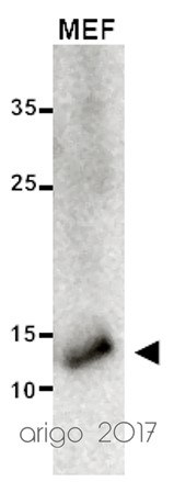
anti-LC3B antibody
Application:WB
Sample:MEF
Sample Loading Amount:20 µg
Primary Antibody Dilution Factor:1:1000
Primary Antibody Incubation Time:overnight
Primary Antibody Incubation Temperature:4 ºC

Average

anti-LC3B antibody
Application:WB
Sample:SW620
Sample Loading Amount:30 µg
Primary Antibody Dilution Factor:1:1000
Primary Antibody Incubation Time:overnight
Primary Antibody Incubation Temperature:4 ºC
文献引用
Phytoagent deoxyelephantopin derivative inhibits triple negative breast cancer cell activity by inducing oxidative stress-mediated paraptosis-like cell death.
WB / Human


