ARG52328
anti-MAP2 antibody
anti-MAP2 antibody for ICC/IF,IHC-Formalin-fixed paraffin-embedded sections,IHC-Frozen sections,Immunohistochemistry (PFA perfusion fixed frozen sections),Western blot and Bovine,Dog,Human,Marmoset,Mouse,Rat,Sheep
Controls and Markers antibody; Neuroscience antibody; Signaling Transduction antibody; Neuron Marker antibody; Mature Neuron Marker antibody; Neurite Marker antibody

概述
| 产品描述 | Chicken Polyclonal antibody recognizes MAP2 |
|---|---|
| 反应物种 | Hu, Ms, Rat, Bov, Dog, Marmoset, Sheep |
| 应用 | ICC/IF, IHC-FoFr , IHC-Fr, IHC-P, WB |
| 宿主 | Chicken |
| 克隆 | Polyclonal |
| 同位型 | IgY |
| 靶点名称 | MAP2 |
| 抗原物种 | Bovine |
| 抗原 | Bovine MAP2 isolated from brain by the GTP microtubule cycling method |
| 偶联标记 | Un-conjugated |
| 別名 | MAP2A; Microtubule-associated protein 2; MAP2C; MAP2B; MAP-2 |
应用说明
| 应用建议 |
|
||||||||||||
|---|---|---|---|---|---|---|---|---|---|---|---|---|---|
| 应用说明 | Specific for the ~ 280k MAP2 protein. * The dilutions indicate recommended starting dilutions and the optimal dilutions or concentrations should be determined by the scientist. |
属性
| 形式 | Liquid |
|---|---|
| 纯化 | Total IgY fraction |
| 缓冲液 | Total IgY fraction in PBS and 10 mM Sodium azide |
| 抗菌剂 | 10 mM Sodium azide |
| 存放说明 | For continuous use, store undiluted antibody at 2-8°C for up to a week. For long-term storage, aliquot and store at -20°C or below. Storage in frost free freezers is not recommended. Avoid repeated freeze/thaw cycles. Suggest spin the vial prior to opening. The antibody solution should be gently mixed before use. |
| 注意事项 | For laboratory research only, not for drug, diagnostic or other use. |
生物信息
| 数据库连接 | |
|---|---|
| 基因名称 | MAP2 |
| 全名 | microtubule associated protein 2 |
| 背景介绍 | Microtubules are 25nm diameter protein rods found in most kinds of eukaryotic cells. They are polymerized from a dimeric subunit made of one a subunit and one b tubulin subunit. Microtubules are associated with a family of proteins called microtubule associated proteins (MAPs), which includes the protein τ (tau) and a group of proteins referred to as MAP1, MAP2, MAP3, MAP4 and MAP5 (Kindler & Gardner 1994). MAP2 is made up of two ~280kDa apparent molecular weight bands referred to as MAP2a and MAP2b. A third lower molecular weight form, usually called MAP2c, corresponds to a pair of protein bands running at ~70kDa on SDS-PAGE gels. All these MAP2 forms are derived from a single gene by alternate transcription, and all share a C-terminal sequence which includes either three or four microtubule binding peptide sequences, which are very similar to those found in the related microtubule binding protein τ (tau). MAP2 isoforms are expressed only in neuronal cells and specifically in the perikarya and dendrites of these cells. MAP2 has been recently shown to be the specific receptor for the neurosteroid pregnenolone (Fontaine-Lenore V. et al., 2006). |
| 产品亮点 | Related Antibody Duos and Panels: ARG30009 NSC and Neuron Marker Antibody Duo (Nestin, MAP2) ARG30301 Neurite Marker Antibody Duo Related products: MAP2 antibodies; MAP2 Duos / Panels; Anti-Chicken IgY secondary antibodies; Related news: 14-3-3η as a promising target for the treatment of Major Depression Disorder Neuronal Development Marker Astrocyte-to-neuron conversion for Parkinson's disease treatment |
| 研究领域 | Controls and Markers antibody; Neuroscience antibody; Signaling Transduction antibody; Neuron Marker antibody; Mature Neuron Marker antibody; Neurite Marker antibody |
| 预测分子量 | 200 kDa |
| 翻译后修饰 | Phosphorylated at serine residues in K-X-G-S motifs by MAP/microtubule affinity-regulating kinase (MARK1 or MARK2), causing detachment from microtubules, and their disassembly (By similarity). Isoform 2 is probably phosphorylated by PKA at Ser-323, Ser-354 and Ser-386 and by FYN at Tyr-67. The interaction with KNDC1 enhances MAP2 threonine phosphorylation (By similarity). |
检测图片 (3) Click the Picture to Zoom In
-

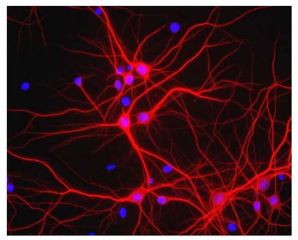
ARG52328 anti-MAP2 antibody ICC/IF image
Immunofluorescence: Mixed neuron/glial cultures. The perikarya and dendrites of neurons are strongly and specifically stained with ARG52328 anti-MAP2 antibody (red). Cell nuclei are visualized with DAPI DNA stain.
-
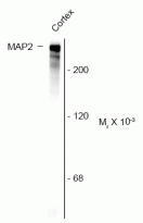
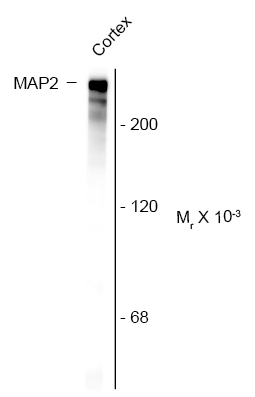
ARG52328 anti-MAP2 antibody WB image
Western blot: Rat cortex lysate stained with ARG52328 anti-MAP2 antibody showing specific immunolabeling of the ~ 280 kDa MAP2 protein.
-
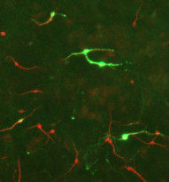
ARG52328 anti-MAP2 antibody ICC/IF image
Immunofluorescence: E17 Rat midbrain mixed neuronal cultures stained with ARG52461 anti-Tyrosine Hydroxylase antibody (green) and ARG52328 anti-MAP2 antibody (red).

客户反馈
Excellent

anti-MAP2 antibody
Application:IHC
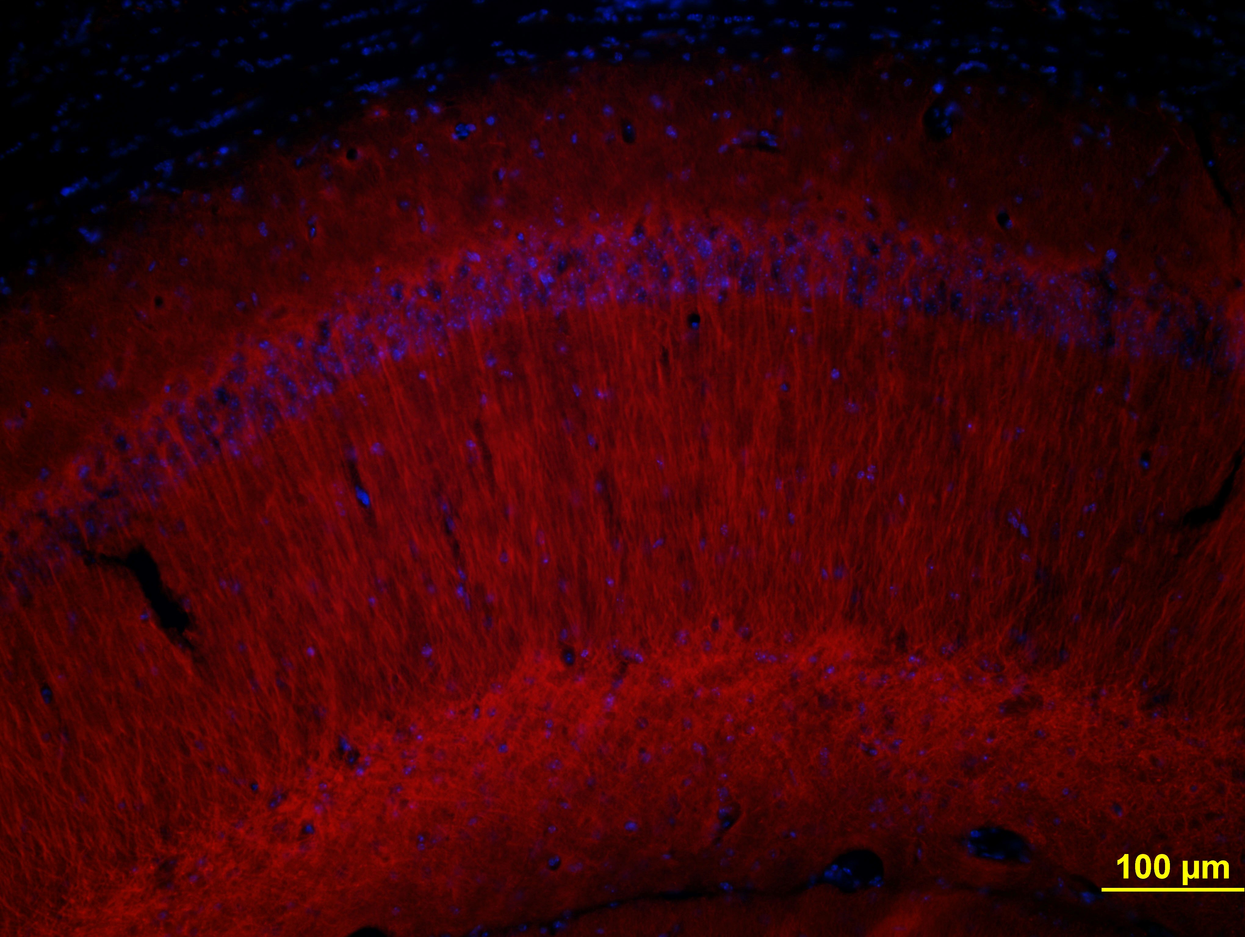
Sample:Mouse brain tissue (hippocampal dentate gyrus)
Sample Preparation Method:Frozen
Fixation Buffer:4% paraformaldehyde
Fixation Time:overnight
Fixation Temperature:4 ºC
Permeabilization Buffer:0.1% TritonX-100
Primary Antibody Dilution Factor:1:500
Primary Antibody Incubation Time:overnight
Primary Antibody Incubation Temperature:4 ºC
Conjugation of Secondary Antibody:Alexa-594

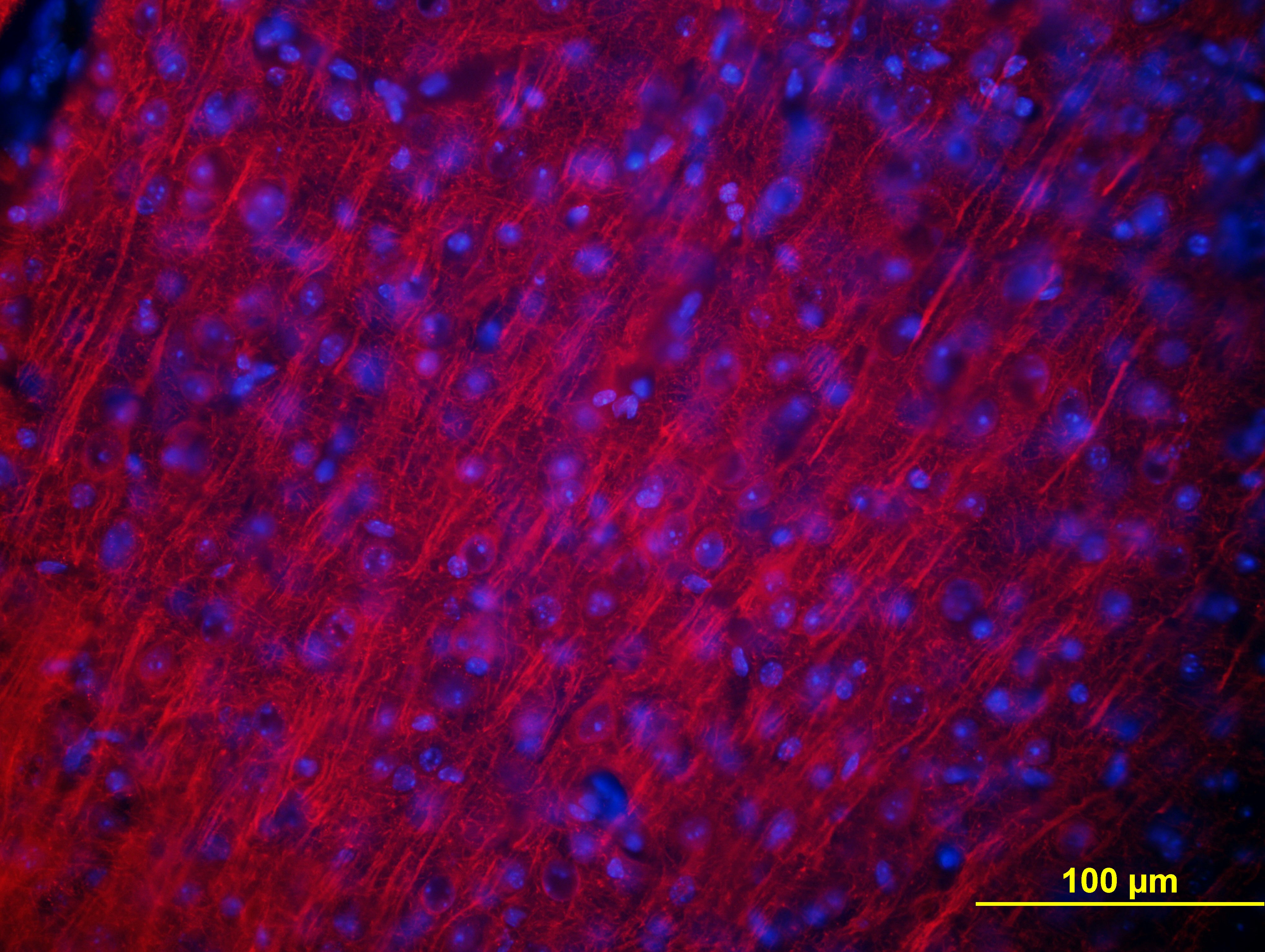
Excellent

anti-MAP2 antibody
Application:IHC
Sample:Mouse brain tissue (hippocampal CA3)
Sample Preparation Method:Frozen
Fixation Buffer:4% paraformaldehyde
Fixation Time:overnight
Fixation Temperature:4 ºC
Permeabilization Buffer:0.1% TritonX-100
Primary Antibody Dilution Factor:1:500
Primary Antibody Incubation Time:overnight
Primary Antibody Incubation Temperature:4 ºC
Conjugation of Secondary Antibody:Alexa-594
Excellent
anti-MAP2 antibody
Application:IHC
Sample:Mouse brain tissue (hippocampal CA1)
Sample Preparation Method:Frozen
Fixation Buffer:4% paraformaldehyde
Fixation Time:overnight
Fixation Temperature:4 ºC
Permeabilization Buffer:0.1% TritonX-100
Primary Antibody Dilution Factor:1:500
Primary Antibody Incubation Time:overnight
Primary Antibody Incubation Temperature:4 ºC
Conjugation of Secondary Antibody:Alexa-594
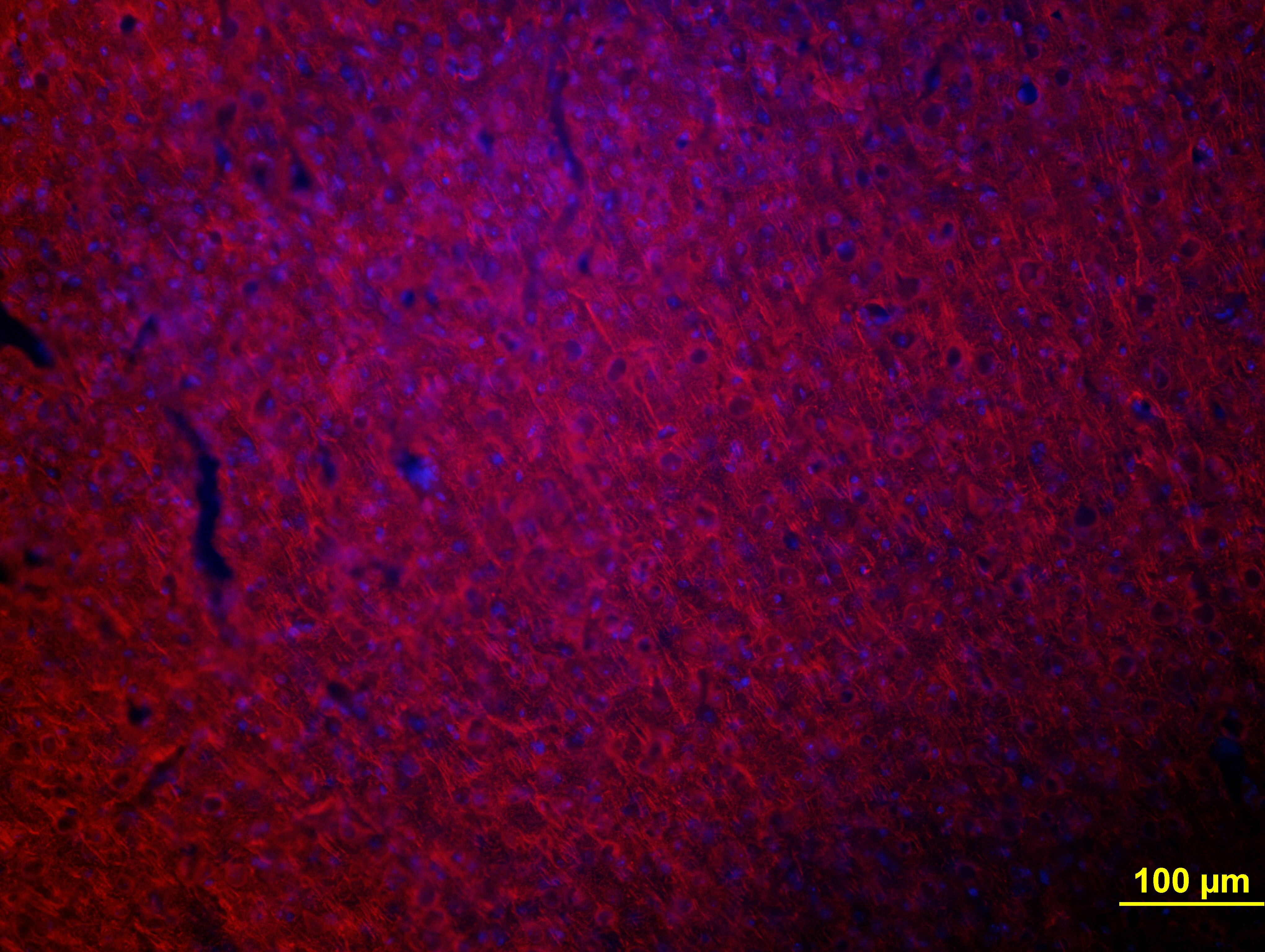
Excellent
anti-MAP2 antibody
Application:IHC
Sample:Mouse brain tissue (cortex)
Sample Preparation Method:Frozen
Fixation Buffer:4% paraformaldehyde
Fixation Time:overnight
Fixation Temperature:4 ºC
Permeabilization Buffer:0.1% TritonX-100
Primary Antibody Dilution Factor:1:500
Primary Antibody Incubation Time:overnight
Primary Antibody Incubation Temperature:4 ºC
Conjugation of Secondary Antibody:Alexa-594
文献引用
Impaired peroxisomal beta-oxidation in microglia triggers oxidative stress and impacts neurons and oligodendrocytes
ICC-IF / Mouse


Magnetic stirring with iron oxide nanospinners accretes neurotoxic Aβ42 oligomers into phagocytic clearable plaques for Alzheimer's disease treatment
ICC/IF / Mouse
Extracellular RNAs-TLR3 signaling contributes to cognitive decline in a mouse model of postoperative cognitive dysfunction.
IHC-Fr / Mouse


