ARG10722
anti-Myelin Basic Protein antibody [7D2]
anti-Myelin Basic Protein antibody [7D2] for ICC/IF,IHC-Frozen sections,Western blot and Human,Mouse,Rat,Cow,Horse,Pig
概述
| 产品描述 | Mouse Monoclonal antibody [7D2] recognizes Myelin Basic Protein |
|---|---|
| 反应物种 | Hu, Ms, Rat, Cow, Hrs, Pig |
| 预测物种 | Chk |
| 应用 | ICC/IF, IHC-Fr, WB |
| 宿主 | Mouse |
| 克隆 | Monoclonal |
| 克隆号 | 7D2 |
| 同位型 | IgG1 |
| 靶点名称 | Myelin Basic Protein |
| 抗原 | Purified myelin basic protein isolated from cow nerve. |
| 偶联标记 | Un-conjugated |
| 別名 | Myelin A1 protein; MBP; Myelin membrane encephalitogenic protein; Myelin basic protein |
应用说明
| 应用建议 |
|
||||||||
|---|---|---|---|---|---|---|---|---|---|
| 应用说明 | * The dilutions indicate recommended starting dilutions and the optimal dilutions or concentrations should be determined by the scientist. |
属性
| 形式 | Liquid |
|---|---|
| 纯化 | Ascites fluid. |
| 缓冲液 | Ascites fluid. |
| 存放说明 | For continuous use, store undiluted antibody at 2-8°C for up to a week. For long-term storage, aliquot and store at -20°C or below. Storage in frost free freezers is not recommended. Avoid repeated freeze/thaw cycles. Suggest spin the vial prior to opening. The antibody solution should be gently mixed before use. |
| 注意事项 | For laboratory research only, not for drug, diagnostic or other use. |
生物信息
| 数据库连接 | |
|---|---|
| 基因名称 | MBP |
| 全名 | myelin basic protein |
| 背景介绍 | The protein encoded by the classic MBP gene is a major constituent of the myelin sheath of oligodendrocytes and Schwann cells in the nervous system. However, MBP-related transcripts are also present in the bone marrow and the immune system. These mRNAs arise from the long MBP gene (otherwise called "Golli-MBP") that contains 3 additional exons located upstream of the classic MBP exons. Alternative splicing from the Golli and the MBP transcription start sites gives rise to 2 sets of MBP-related transcripts and gene products. The Golli mRNAs contain 3 exons unique to Golli-MBP, spliced in-frame to 1 or more MBP exons. They encode hybrid proteins that have N-terminal Golli aa sequence linked to MBP aa sequence. The second family of transcripts contain only MBP exons and produce the well characterized myelin basic proteins. This complex gene structure is conserved among species suggesting that the MBP transcription unit is an integral part of the Golli transcription unit and that this arrangement is important for the function and/or regulation of these genes. [provided by RefSeq, Jul 2008] |
| 生物功能 | The classic group of MBP isoforms (isoform 4-isoform 14) are with PLP the most abundant protein components of the myelin membrane in the CNS. They have a role in both its formation and stabilization. The smaller isoforms might have an important role in remyelination of denuded axons in multiple sclerosis. The non-classic group of MBP isoforms (isoform 1-isoform 3/Golli-MBPs) may preferentially have a role in the early developing brain long before myelination, maybe as components of transcriptional complexes, and may also be involved in signaling pathways in T-cells and neural cells. Differential splicing events combined with optional post-translational modifications give a wide spectrum of isomers, with each of them potentially having a specialized function. Induces T-cell proliferation. [UniProt] |
| 预测分子量 | 33 kDa |
| 翻译后修饰 | Several charge isomers of MBP; C1 (the most cationic, least modified, and most abundant form), C2, C3, C4, C5, C6, C7, C8-A and C8-B (the least cationic form); are produced as a result of optional PTM, such as phosphorylation, deamidation of glutamine or asparagine, arginine citrullination and methylation. C8-A and C8-B contain each two mass isoforms termed C8-A(H), C8-A(L), C8-B(H) and C8-B(L), (H) standing for higher and (L) for lower molecular weight. C3, C4 and C5 are phosphorylated. The ratio of methylated arginine residues decreases during aging, making the protein more cationic. The N-terminal alanine is acetylated (isoform 3, isoform 4, isoform 5 and isoform 6). Arg-241 was found to be 6% monomethylated and 60% symmetrically dimethylated. Phosphorylated by TAOK2, VRK2, MAPK11, MAPK12, MAPK14 and MINK1. |
检测图片 (5) Click the Picture to Zoom In
-

ARG10722 anti-Myelin Basic Protein antibody [7D2] ICC/IF image
Immunocytochemistry: Rat mixed neuron / glial cultures stained with ARG10722 anti-Myelin Basic Protein antibody [7D2] (red) and co-stained with chicken antibody to neurofilament NF-L (green). Blue is a DNA stain. Note that the MBP antibody stains an oligodendrocyte and some membrane shed from this cell. Other cells in the field include neurons, astrocytes, microglia and fibroblasts, all of which are completely negative for MBP, though the neuronal processes can be seen with the NF-L antibody.
-

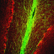
ARG10722 anti-Myelin Basic Protein antibody [7D2] IHC-Fr image
Immunohistochemistry: Frozen section of Rat brain hippocampus stained with ARG10722 anti-Myelin Basic Protein antibody [7D2] (green) at 1:5000 dilution and costained with ARG10724 anti-Neurofilament NF-M antibody (red) at 1:2000 dilution.
The Myelin Basic Protein antibody stains myelin sheathes around axons, while the NF-M antibody labels dendrites and axons of neuronal cells.
-
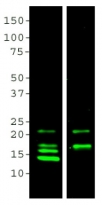
ARG10722 anti-Myelin Basic Protein antibody [7D2] WB image
Western blot: 20 µg of crude Rat brain homogenate were stained with two MBP antibodies; Clone 7G7 (lane 1) at 1:5000 and ARG10722 anti-Myelin Basic Protein antibody [7D2] (lane 2) at 1:5000. MCA-7D2 binds the largest 21.5 kDa and 18.5 kDa transcripts preferentially, while monoclonal MCA-7G7 bind all four transcripts: 21.5 kDa, 18.5 kDa, 17 kDa and 14 kDa.
-
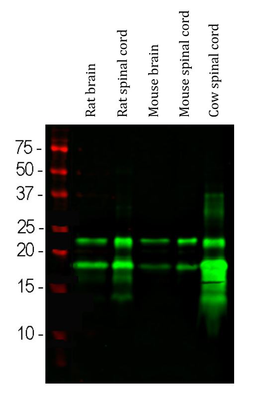
ARG10722 anti-Myelin Basic Protein antibody [7D2] WB image
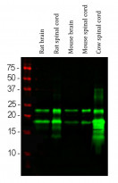
Western blot: Rat brain, Rat spinal cord, Mouse brain, Mouse spinal cord and Cow spinal cord lysates stained with ARG10722 anti-Myelin Basic Protein antibody [7D2] (green) at 1:10000 dilution.
-
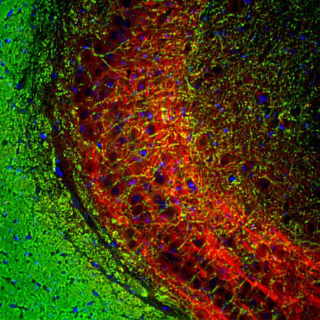
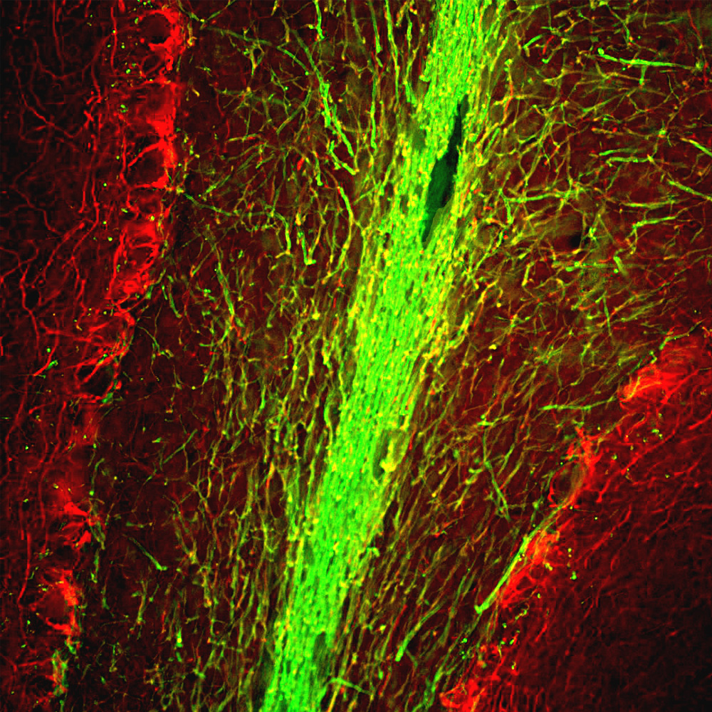
ARG10722 anti-Myelin Basic Protein antibody [7D2] IHC-Fr image
Immunohistochemistry: Frozen section of Rat brain cerebellum stained with ARG10722 anti-Myelin Basic Protein antibody [7D2] (green) at 1:5000 dilution and costained with ARG10724 anti-Neurofilament NF-M antibody (red) at 1:2000 dilution.
The Myelin Basic Protein antibody stains myelin sheathes around axons, while the NF-M antibody labels dendrites and axons of neuronal cells.


文献引用
Activin A rescues preterm brain injury through a novel Noggin/BMP4/Id2 signaling pathway
WB, IHC-Fr / Rat


