ARG42026
anti-SQSTM1 / p62 antibody [3H11]
anti-SQSTM1 / p62 antibody [3H11] for Flow cytometry,ICC/IF,IHC-Formalin-fixed paraffin-embedded sections,Western blot and Human,Mouse,Rat
Cancer antibody; Cell Biology and Cellular Response antibody; Cell Death antibody; Gene Regulation antibody; Metabolism antibody; Signaling Transduction antibody; Autophagy Study antibody
概述
| 产品描述 | Mouse Monoclonal antibody [3H11] recognizes SQSTM1 / p62 |
|---|---|
| 反应物种 | Hu, Ms, Rat |
| 应用 | FACS, ICC/IF, IHC-P, WB |
| 宿主 | Mouse |
| 克隆 | Monoclonal |
| 克隆号 | 3H11 |
| 同位型 | IgG2a |
| 靶点名称 | SQSTM1 / p62 |
| 抗原物种 | Human |
| 抗原 | Synthetic peptide corresponding to aa. 69-96 of Human SQSTM1 / p62. (DEDGDLVAFSSDEELTMAMSYVKDDIFR) |
| 偶联标记 | Un-conjugated |
| 別名 | FTDALS3; p60; p62; EBI3-associated protein of 60 kDa; p62B; Sequestosome-1; Ubiquitin-binding protein p62; PDB3; OSIL; Phosphotyrosine-independent ligand for the Lck SH2 domain of 62 kDa; EBIAP; A170; ZIP3 |
应用说明
| 应用建议 |
|
||||||||||
|---|---|---|---|---|---|---|---|---|---|---|---|
| 应用说明 | IHC-P: Antigen Retrieval: Heat mediation was performed in Citrate buffer (pH 6.0, epitope retrieval solution) for 20 min. * The dilutions indicate recommended starting dilutions and the optimal dilutions or concentrations should be determined by the scientist. |
||||||||||
| 实际分子量 | ~ 62 kDa |
属性
| 形式 | Liquid |
|---|---|
| 纯化 | Affinity purification with immunogen. |
| 缓冲液 | 0.2% Na2HPO4, 0.9% NaCl, 0.05% Sodium azide and 4% Trehalose. |
| 抗菌剂 | 0.05% Sodium azide |
| 稳定剂 | 4% Trehalose |
| 浓度 | 0.5 mg/ml |
| 存放说明 | For continuous use, store undiluted antibody at 2-8°C for up to a week. For long-term storage, aliquot and store at -20°C or below. Storage in frost free freezers is not recommended. Avoid repeated freeze/thaw cycles. Suggest spin the vial prior to opening. The antibody solution should be gently mixed before use. |
| 注意事项 | For laboratory research only, not for drug, diagnostic or other use. |
生物信息
| 数据库连接 | |
|---|---|
| 基因名称 | SQSTM1 |
| 全名 | sequestosome 1 |
| 背景介绍 | This gene encodes a multifunctional protein that binds ubiquitin and regulates activation of the nuclear factor kappa-B (NF-kB) signaling pathway. The protein functions as a scaffolding/adaptor protein in concert with TNF receptor-associated factor 6 to mediate activation of NF-kB in response to upstream signals. Alternatively spliced transcript variants encoding either the same or different isoforms have been identified for this gene. Mutations in this gene result in sporadic and familial Paget disease of bone. [provided by RefSeq, Mar 2009] |
| 生物功能 | Autophagy receptor that interacts directly with both the cargo to become degraded and an autophagy modifier of the MAP1 LC3 family. Required both for the formation and autophagic degradation of polyubiquitin-containing bodies, called ALIS (aggresome-like induced structures) and links ALIS to the autophagic machinery. Involved in midbody ring degradation. May regulate the activation of NFKB1 by TNF-alpha, nerve growth factor (NGF) and interleukin-1. May play a role in titin/TTN downstream signaling in muscle cells. May regulate signaling cascades through ubiquitination. Adapter that mediates the interaction between TRAF6 and CYLD (By similarity). May be involved in cell differentiation, apoptosis, immune response and regulation of K(+) channels. [UniProt] |
| 细胞定位 | Cytoplasm, cytosol. Late endosome. Lysosome. Cytoplasmic vesicle, autophagosome. Nucleus. Endoplasmic reticulum. Nucleus, PML body. Cytoplasm, myofibril, sarcomere. [UniProt] |
| 产品亮点 | Related products: SQSTM1 antibodies; SQSTM1 Duos / Panels; Anti-Mouse IgG secondary antibodies; Related news: Keap1-Nrf2-ARE antibody panel is launched Therapeutic strategies against PDAC |
| 研究领域 | Cancer antibody; Cell Biology and Cellular Response antibody; Cell Death antibody; Gene Regulation antibody; Metabolism antibody; Signaling Transduction antibody; Autophagy Study antibody |
| 预测分子量 | 48 kDa |
| 翻译后修饰 | Phosphorylated. May be phosphorylated by PRKCZ (By similarity). Phosphorylated in vitro by TTN. Phosphorylation at Ser-403 by ULK1 is stimulated by SESN2 (PubMed:25040165). Ubiquitinated by RNF26: ubiquitinated SQSTM1 attracts specific vesicle-associated adapters, forming a molecular bridge that restrains cognate vesicles in the perinuclear region and organizes the endosomal pathway for efficient cargo transport (PubMed:27368102). Deubiquitination by USP15 releases target vesicles for fast transport into the cell periphery (PubMed:27368102). [UniProt] |
检测图片 (10) Click the Picture to Zoom In
-

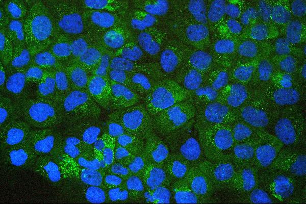
ARG42026 anti-SQSTM1 / p62 antibody [3H11] ICC/IF image
Immunofluorescence: A431 cells stained with ARG42026 anti-SQSTM1 / p62 antibody [3H11] (green) at 2 µg/ml dilution, overnight at 4°C. DAPI (blue) for nuclear staining.
-

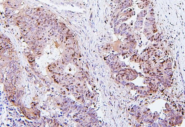
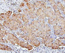
ARG42026 anti-SQSTM1 / p62 antibody [3H11] IHC-P image
Immunohistochemistry: Paraffin-embedded Human colon cancer tissue. Antigen Retrieval: Heat mediation was performed in Citrate buffer (pH 6.0, epitope retrieval solution) for 20 min. The tissue section was blocked with 10% goat serum. The tissue section was then stained with ARG42026 anti-SQSTM1 / p62 antibody [3H11] at 1 µg/ml dilution, overnight at 4°C.
-
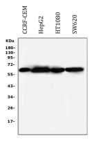
ARG42026 anti-SQSTM1 / p62 antibody [3H11] WB image
Western blot: 50 µg of samples under reducing conditions. CCRF-CEM, HepG2, HT1080 and SW620 whole cell lysates stained with ARG42026 anti-SQSTM1 / p62 antibody [3H11] at 0.5 µg/ml dilution, overnight at 4°C.
-
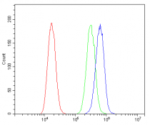
ARG42026 anti-SQSTM1 / p62 antibody [3H11] FACS image
Flow Cytometry: A549 cells were blocked with 10% normal goat serum and then stained with ARG42026 anti-SQSTM1 / p62 antibody [3H11] (blue) at 1 µg/10^6 cells for 30 min at 20°C, followed by incubation with DyLight®488 labelled secondary antibody. Isotype control antibody (green) was Mouse IgG (1 µg/10^6 cells) used under the same conditions. Unlabelled sample (red) was also used as a control.
-
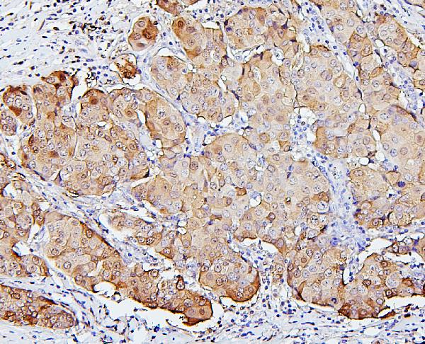
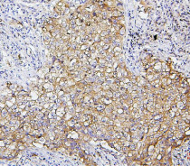
ARG42026 anti-SQSTM1 / p62 antibody [3H11] IHC-P image
Immunohistochemistry: Paraffin-embedded Human mammary cancer tissue. Antigen Retrieval: Heat mediation was performed in Citrate buffer (pH 6.0, epitope retrieval solution) for 20 min. The tissue section was blocked with 10% goat serum. The tissue section was then stained with ARG42026 anti-SQSTM1 / p62 antibody [3H11] at 1 µg/ml dilution, overnight at 4°C.
-
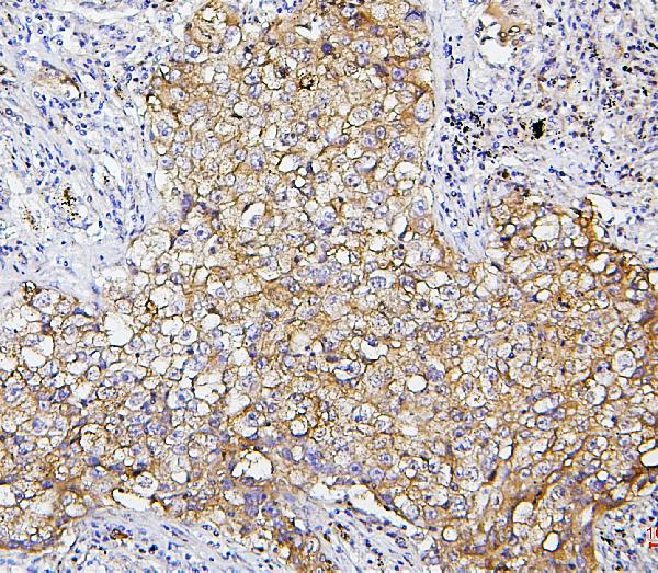
ARG42026 anti-SQSTM1 / p62 antibody [3H11] IHC-P image
Immunohistochemistry: Paraffin-embedded Human lung cancer tissue. Antigen Retrieval: Heat mediation was performed in Citrate buffer (pH 6.0, epitope retrieval solution) for 20 min. The tissue section was blocked with 10% goat serum. The tissue section was then stained with ARG42026 anti-SQSTM1 / p62 antibody [3H11] at 1 µg/ml dilution, overnight at 4°C.
-
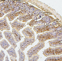
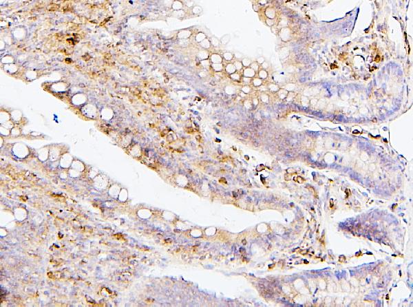
ARG42026 anti-SQSTM1 / p62 antibody [3H11] IHC-P image
Immunohistochemistry: Paraffin-embedded Mouse intestine tissue. Antigen Retrieval: Heat mediation was performed in Citrate buffer (pH 6.0, epitope retrieval solution) for 20 min. The tissue section was blocked with 10% goat serum. The tissue section was then stained with ARG42026 anti-SQSTM1 / p62 antibody [3H11] at 1 µg/ml dilution, overnight at 4°C.
-
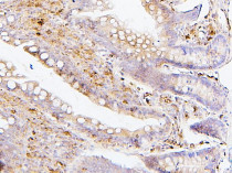
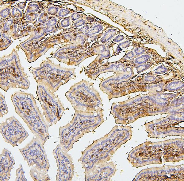
ARG42026 anti-SQSTM1 / p62 antibody [3H11] IHC-P image
Immunohistochemistry: Paraffin-embedded Rat intestine tissue. Antigen Retrieval: Heat mediation was performed in Citrate buffer (pH 6.0, epitope retrieval solution) for 20 min. The tissue section was blocked with 10% goat serum. The tissue section was then stained with ARG42026 anti-SQSTM1 / p62 antibody [3H11] at 1 µg/ml dilution, overnight at 4°C.
-

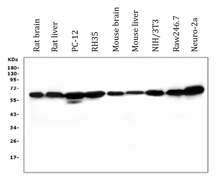
ARG42026 anti-SQSTM1 / p62 antibody [3H11] WB image
Western blot: 50 µg of samples under reducing conditions. Rat brain, Rat liver, PC-12, RH35, Mouse brain, Mouse liver, NIH/3T3, Raw246.7 and Neuro-2a whole cell lysates stained with ARG42026 anti-SQSTM1 / p62 antibody [3H11] at 0.5 µg/ml dilution, overnight at 4°C.
-
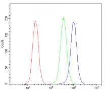
ARG42026 anti-SQSTM1 / p62 antibody [3H11] FACS image
Flow Cytometry: PC-3 cells were blocked with 10% normal goat serum and then stained with ARG42026 anti-SQSTM1 / p62 antibody [3H11] (blue) at 1 µg/10^6 cells for 30 min at 20°C, followed by incubation with DyLight®488 labelled secondary antibody. Isotype control antibody (green) was Mouse IgG (1 µg/10^6 cells) used under the same conditions. Unlabelled sample (red) was also used as a control.