ARG52466
anti-UCHL1 / PGP9.5 antibody
anti-UCHL1 / PGP9.5 antibody for ICC/IF,Western blot and Human,Mouse,Rat
Cell Biology and Cellular Response antibody; Gene Regulation antibody; Neuroscience antibody
概述
| 产品描述 | Chicken Polyclonal antibody recognizes UCHL1 / PGP9.5 |
|---|---|
| 反应物种 | Hu, Ms, Rat |
| 预测物种 | Mamm |
| 应用 | ICC/IF, WB |
| 宿主 | Chicken |
| 克隆 | Polyclonal |
| 同位型 | IgY |
| 靶点名称 | UCHL1 / PGP9.5 |
| 抗原物种 | Human |
| 抗原 | Recombinant full length human UCHL1 purified from E. coli |
| 偶联标记 | Un-conjugated |
| 別名 | PGP95; UCH-L1; PGP9.5; PARK5; Ubiquitin thioesterase L1; HEL-117; Neuron cytoplasmic protein 9.5; Uch-L1; EC 6.-.-.-; PGP 9.5; Ubiquitin carboxyl-terminal hydrolase isozyme L1; NDGOA; EC 3.4.19.12 |
应用说明
| 应用建议 |
|
||||||
|---|---|---|---|---|---|---|---|
| 应用说明 | Specific for the ~24kDa UCHL1 protein. * The dilutions indicate recommended starting dilutions and the optimal dilutions or concentrations should be determined by the scientist. |
属性
| 形式 | Liquid |
|---|---|
| 纯化 | Total IgY fraction |
| 缓冲液 | Total IgY fraction in PBS and 10 mM Sodium azide |
| 抗菌剂 | 10 mM Sodium azide |
| 存放说明 | For continuous use, store undiluted antibody at 2-8°C for up to a week. For long-term storage, aliquot and store at -20°C or below. Storage in frost free freezers is not recommended. Avoid repeated freeze/thaw cycles. Suggest spin the vial prior to opening. The antibody solution should be gently mixed before use. |
| 注意事项 | For laboratory research only, not for drug, diagnostic or other use. |
生物信息
| 数据库连接 | |
|---|---|
| 基因名称 | UCHL1 |
| 全名 | ubiquitin carboxyl-terminal esterase L1 (ubiquitin thiolesterase) |
| 背景介绍 | Ubiquitin C-terminal hydrolase 1 (UCHL1) is also known as ubiquitin carboxyl esterase L1, ubiquitin thiolesterase, neuron-specific protein PGP9.5 and Park5. It was originally identified as a major component of the neuronal cytoplasm from 2-dimensional gel analysis of brain tissues, and was given the name PGP9.5 . It was later found that ubiquitin C-terminal hydrolase enzyme activity was associated with the PGP9.5 protein . The ubiquitin C-terminal hydrolases cleave ubiquitin from other molecules. Regulation of the ubiquitin pathway is very important and many disease states are associated with defects in this pathway. Genetic knockout of UCHL1 in mice results in a motor neuron degeneration similar to the spontaneous gracile axonal dystrophy (gad) mutant mice . Point mutations in the UCHL1 gene are associated with some forms of human Parkinson's disease . Since UCHL1 is heavily expressed in neurons, it is released in large amounts following injury or degeneration, so the detection of UCHL1 in CSF and other bodily fluids can be used as a biomarker. |
| 研究领域 | Cell Biology and Cellular Response antibody; Gene Regulation antibody; Neuroscience antibody |
| 预测分子量 | 25 kDa |
| 翻译后修饰 | O-glycosylated. |
检测图片 (4) Click the Picture to Zoom In
-
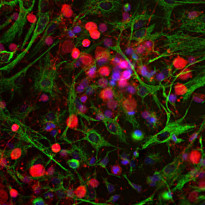
ARG52466 anti-UCHL1 / PGP9.5 antibody ICC/IF image
Immunofluorescence: Cortical neuron-glial cell culture from E20 Rat stained with ARG52466 anti-UCHL1 / PGP9.5 antibody (red) at 1:500 dilution, and costained with anti-Vimentin antibody (green) at 1:2000 dilution. DAPI (blue) for nuclear staining.
The UCHL1 antibody produces strong staining of the cell body and dendrites in neurons. The Vimentin antibody stains intermediate filaments in fibroblastic and developing glial cells.
-
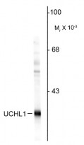
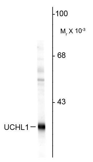
ARG52466 anti-UCHL1 / PGP9.5 antibody WB image
Western blot: Rat hippocampal homogenate stained with ARG52466 anti-UCHL1 / PGP9.5 antibody showing specific immunolabeling of the ~ 24k UCHL1 protein.
-
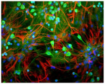
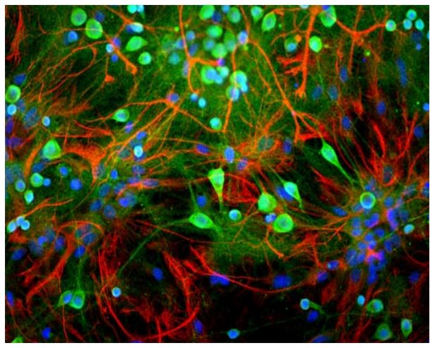
ARG52466 anti-UCHL1 / PGP9.5 antibody ICC/IF image
Immunofluorescence: Rat mixed neuron/glial cultures stained with ARG52466 anti-UCHL1 / PGP9.5 antibody (green) and ARG52312 rabbit anti-GFAP antibody (red). Blue is a DNA stain.
-
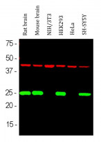
ARG52466 anti-UCHL1 / PGP9.5 antibody WB image
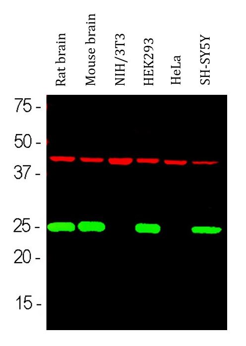
Western blot: Rat brain, Mouse brain, NIH/3T3, HEK293, HeLa and SH-SY5Y cell lysates stained with ARG52466 anti-UCHL1 / PGP9.5 antibody (green) at 1:2000 dilution and anti-Actin antibody (red) at 1:1000 dilution.
The single band at 24 kDa mark corresponds to UCHL1 protein which is detectable in CNS extracts and lysates of cells with neuronal properties but not in lysates of HeLa, NIH/3T3 and other non-neuronal cells. Actin is detected with apparent molecular weight of 42 kDa and provides an loading control.