ARG10728
anti-Ubiquilin 2 antibody [6H9]
anti-Ubiquilin 2 antibody [6H9] for ICC/IF,IHC-Frozen sections,Western blot and Human,Mouse,Rat
概述
| 产品描述 | Mouse Monoclonal antibody [6H9] recognizes Ubiquilin 2 |
|---|---|
| 反应物种 | Hu, Ms, Rat |
| 应用 | ICC/IF, IHC-Fr, WB |
| 特异性 | This antibody highly selective the Ubiquilin 2, little cross to Ubiquilin 1. |
| 宿主 | Mouse |
| 克隆 | Monoclonal |
| 克隆号 | 6H9 |
| 同位型 | IgG1 |
| 靶点名称 | Ubiquilin 2 |
| 抗原物种 | Human |
| 抗原 | Human Ubiquilin 2 expressed in and purified from E. coli. |
| 偶联标记 | Un-conjugated |
| 別名 | PLIC-2; Ubiquitin-like product Chap1/Dsk2; HRIHFB2157; Chap1; DSK2 homolog; PLIC2; ALS15; DSK2; CHAP1; Protein linking IAP with cytoskeleton 2; N4BP4; Ubiquilin-2; hPLIC-2 |
应用说明
| 应用建议 |
|
||||||||
|---|---|---|---|---|---|---|---|---|---|
| 应用说明 | * The dilutions indicate recommended starting dilutions and the optimal dilutions or concentrations should be determined by the scientist. |
属性
| 形式 | Liquid |
|---|---|
| 纯化 | Affinity purification. |
| 缓冲液 | PBS and 50% Glycerol. |
| 稳定剂 | 50% Glycerol |
| 浓度 | 1 mg/ml |
| 存放说明 | For continuous use, store undiluted antibody at 2-8°C for up to a week. For long-term storage, aliquot and store at -20°C. Storage in frost free freezers is not recommended. Avoid repeated freeze/thaw cycles. Suggest spin the vial prior to opening. The antibody solution should be gently mixed before use. |
| 注意事项 | For laboratory research only, not for drug, diagnostic or other use. |
生物信息
| 数据库连接 | |
|---|---|
| 基因名称 | UBQLN2 |
| 全名 | ubiquilin 2 |
| 背景介绍 | This gene encodes an ubiquitin-like protein (ubiquilin) that shares high degree of similarity with related products in yeast, rat and frog. Ubiquilins contain a N-terminal ubiquitin-like domain and a C-terminal ubiquitin-associated domain. They physically associate with both proteasomes and ubiquitin ligases; and thus, are thought to functionally link the ubiquitination machinery to the proteasome to affect in vivo protein degradation. This ubiquilin has also been shown to bind the ATPase domain of the Hsp70-like Stch protein. [provided by RefSeq, Oct 2009] |
| 生物功能 | Plays an important role in the regulation of different protein degradation mechanisms and pathways including ubiquitin-proteasome system (UPS), autophagy and the endoplasmic reticulum-associated protein degradation (ERAD) pathway. Mediates the proteasomal targeting of misfolded or accumulated proteins for degradation by binding (via UBA domain) to their polyubiquitin chains and by interacting (via ubiquitin-like domain) with the subunits of the proteasome. Plays a role in the ERAD pathway via its interaction with ER-localized proteins FAF2/UBXD8 and HERPUD1 and may form a link between the polyubiquitinated ERAD substrates and the proteasome. Involved in the regulation of macroautophagy and autophagosome formation; required for maturation of autophagy-related protein LC3 from the cytosolic form LC3-I to the membrane-bound form LC3-II and may assist in the maturation of autophagosomes to autolysosomes by mediating autophagosome-lysosome fusion. Negatively regulates the endocytosis of GPCR receptors: AVPR2 and ADRB2, by specifically reducing the rate at which receptor-arrestin complexes concentrate in clathrin-coated pits (CCPs). [UniProt] |
| 预测分子量 | 66 kDa |
| 翻译后修饰 | Degraded during macroautophagy. |
检测图片 (5) Click the Picture to Zoom In
-
ARG10728 anti-Ubiquilin 2 antibody [6H9] ICC/IF image
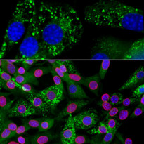
Immunofluorescence: NIH/3T3 cells stained with ARG10728 anti-Ubiquilin 2 antibody [6H9] (green) at 1:1000 dilution and costained with ARG10685 anti-Lamin A + C antibody (red) at 1:5000 dilution. DAPI (blue) for nuclear staining.
The cells were treated with 50 µM of chloroquine, an inhibitor of autophagy, for 16 hours prior to staining. Clone 6H9 antibody reveals punctate staining of ubiquilin 2 protein accumulated in lysosomes in the cytoplasm, while the Lamin A + C antibody stains the nuclear lamina.
-

ARG10728 anti-Ubiquilin 2 antibody [6H9] ICC/IF image
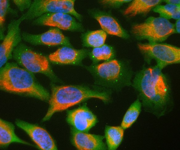
Immunocytochemistry: HeLa cell cultures stained with ARG10728 anti-Ubiquilin 2 antibody [6H9] (green) and co-stained with chicken polyclonal antibody to vimentin (red). In most individual cells, ubiquilin 2 is present diffusely in the cytoplasm of cells, though some cells show enrichment of the protein in spherical autophagosome-like structure.
-
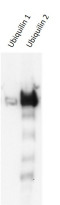
ARG10728 anti-Ubiquilin 2 antibody [6H9] WB image
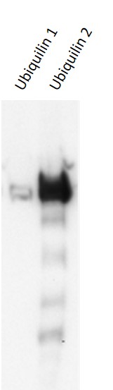
Western blot: Ubiquilin 1 and Ubiquilin 2 recombinant stained with ARG10728 anti-Ubiquilin 2 antibody [6H9].
-
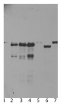
ARG10728 anti-Ubiquilin 2 antibody [6H9] WB image
Western blot: 1) Untransfected primary Mouse neuron and glia cell cultures, 2) the same cells transduced with Human ubiquilin 2 wild type, 3) with ubiquilin 2 P506T mutant, 4) with ubiquilin 2 P497S mutant, and 5) with enhanced GFP control; 6) HeLa cells, and 7) 3T3 cells stained with ARG10728 anti-Ubiquilin 2 antibody [6H9]. In primary mouse neuron and glia cell culture, endogenous ubiquilin 2 appears as a weak band at 68 kDa in all transduced and non-transduced cells, indicating low endogenous expression of mouse ubiquilin 2. Strong bands are seen in the cells transduced with Human wild type or mutant ubiquilin 2. Small proteins run at 50 kDa in these cells are the fragments of ubiquilin 2. Note, ubiquilin 2 run at ~66 kDa in Human HeLa cells and 68 kDa in rodent 3T3 cells.
-
ARG10728 anti-Ubiquilin 2 antibody [6H9] WB image
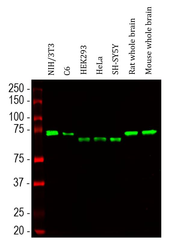
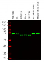
Western blot: NIH/3T3, C6, HEK293, HeLa, SH-SY5Y, Rat whole brain and Mouse whole brain lysates stained with ARG10728 anti-Ubiquilin 2 antibody [6H9] (green) at 1:1000 dilution.